Reduced Acquisition Time [18F]GE-180 PET Scanning Protocol Replaces Gold-Standard Dynamic Acquisition in a Mouse Ischemic Stroke Model
2022.02.10.
Artem Zatcepin ez al., Frontiers in Medicine, 2022
Summary
Stroke is the second leading cause of death worldwide and one of the leading causes of disability. Treatment dedicated to improving post-stroke recovery remains unresolved. One of potential targets for such treatment is post-stroke neuroinflammation due to its long-lasting nature and the presence of multiple effectors that can be targeted. Yet, none of the so far conducted trials testing immunomodulatory therapies have been able to consistently demonstrate a beneficial function of such approaches on post-stroke recovery. Even though the influence of the neuroinflammatory response to stroke outcome in humans is yet to be determined, individual measures of neuroinflammation can assist in patient selection for immune interventions.
Neuroinflammation can be assessed through monitoring of microglia, the brain resident innate immune cells. Microglia activation is strongly correlated to the 18 kDa translocator protein (TSPO) expression level on the outer membrane of microglial mitochondria. However, it should be mentioned that microglia are not the only cell type expressing TSPO in the brain under disease conditions. For instance, activated astrocytes significantly contribute to the overall TSPO signal, as shown in the literature for rats with traumatic brain injury and for patients with multiple sclerosis. Multiple PET tracers targeting TSPO have been developed over the years. First-generation TSPO tracers, such as [11C]-I-PK11195 and [11C]-AC-5216, have low signal-to-noise ratio and high off-target binding, which prevented them from routine use. Second-generation TSPO tracers include flutriciclamide ([18F]GE-180), a tracer that has higher specific binding and improved signal-to-noise ratio.
Benchmark parameters of a microglia PET quantification protocol with [18F]GE-180 consist of a 0–90 min post-injection (p.i.) acquisition, continuous arterial blood sampling in the first 15 min, and measurement of the tracer metabolites in the arterial blood. These data are required to estimate quantitative indices such as total distribution volume (VT) of [18F]GE-180 using the 2-tissue compartment model with reversible binding or the Logan plot. However, it is not feasible to implement such a protocol in most clinical settings due to the complexity of arterial blood sampling, reduced patient comfort, and increased workload for a nuclear medicine department. In small animal studies, especially in longitudinal measurements, the arterial blood sampling is even more challenging, as, due to the limited amount of blood, it requires an external arteriovenous shunt to be installed.
Therefore, it is desirable to establish a simplified quantification approach that would yield a robust TSPO expression estimate without the need for the long PET acquisition and arterial blood sampling. In an experimental stroke model, the authors investigated the relationship between the VT ratio (DVR) in the lesion area and the normal cortex tissue calculated from a long 0–90 min p.i. [18F]GE-180 PET scan by using Logan plot with an image-derived blood input function (IDIF) and the standardized uptake value ratio (SUVR) in the same regions estimated from one of the late 10 min time frames. The authors then established and validated a late 30 min time frame for simplified [18F]GE-180 quantification.
Results from nanoScan PET/MRI 3T
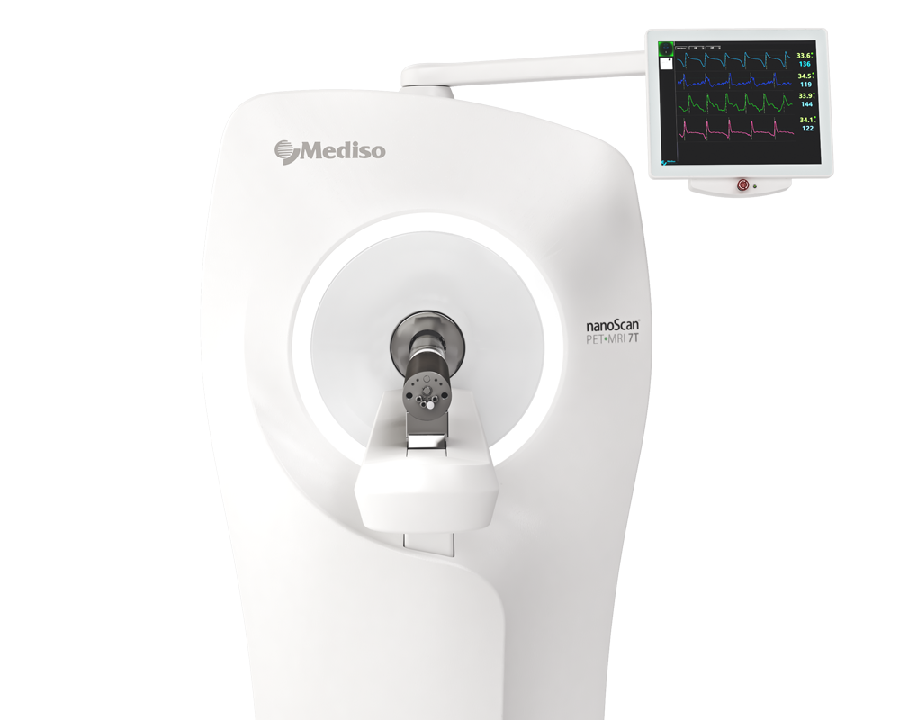
For the imaging studies, The mice were scanned at 7, 28, 84, and 168 days after the photothrombotic stroke (PT) induction using the nanoScan PET/MRI 3T scanner with a single-mouse imaging chamber. The mice received an intravenous injection of 18.0 ± 2.1 MBq [18F]GE-180 through the tail vein. For the dynamic PET imaging (3 PT and 3 sham mice), acquisition was performed from 0 to 90 min p.i. (analysis cohort). For the static PET imaging, (3 PT and 3 sham mice), the list-mode data were acquired at 60-90 min p.i. (validation cohort). A 15-min anatomical T1 MR scan was performed at 30 min after [18F]GE-180 injection for the validation cohort (static imaging) and after 90 min for the analysis cohort (head receive coil, matrix size 96 × 96 × 30, voxel size 0.21 × 0.24 × 0.65 mm3, repetition time 677 ms, echo time 28.56 ms, flip angle 90°). The PET field of view (FOV) included the whole mouse, while the MR FOV covered the mouse head only. The T1 image was then used to create a body-air material map for the attenuation correction of the PET data. The authors reconstructed the PET list-mode data within a 400–600 keV energy window using the Tera-Tomo 3D iterative algorithm with the following parameters: matrix size 55 × 62 × 187 mm3, voxel size 0.3 × 0.3 × 0.3 mm3, 8 iterations, 6 subsets. When acquired dynamically (0–90 min p.i. acquisitions), the list-mode data were binned into 25 frames (6 x 10, 2 x 30 s, 3 x 1, 5 x 2, 5 x 5, 5 x 10 min). Decay and random correction were applied.
Fig 2. shows the time development of PT in a single mouse demonstrated by [18F]GE-180 PET/MR. Timepoints after the surgery: (A) 7 days, (B) 28 days, (C) 84 days, (D) 168 days. VOIs shown in the image: white—cerebellum, black—cerebellar white matter. The location of the sagittal slice on a template MR image is shown on the top right.
Fig. 3. Factor analysis-based PVE correction. (1A)—original dynamic scan (late frame is shown); (1B)—vena cava region; (2A)—factor curves; (2B–E)—factor images; (3A)—blood factor curve f1(t); (3B)—blood factor image a1(i); (4)—spill-in corrected dynamic image S∗iSi*(t) (early frame is shown). The images were used for image-derived blood input function (IDIF) generation for the Logan plot.

The authors examined the late time frames (40–50, 50–60, 60–70, 70–80, and 80–90 min p.i.) from the dynamic [18F]GE-180 PET scans (analysis cohort) to determine which frames yield the SUVR values that approximate the DVR values in the PT VOI best. In this part of the study, the authors performed linear fitting on the combined data (PT and sham). The fit lines of the 60–70, 70–80, and 80–90 min p.i. frames had the slope value closest to one (0.81, 0.91, and 0.96, respectively) and only slightly under- or overestimated the line of identity, which makes them the best approximations among the considered frames. 40−50 and 50–60 min frames significantly underestimated DVR (p = 0.002/p = 0.01).
Based on this finding, the authors then reconstructed the list-mode data from 60 to 90 min p.i. and plotted the corresponding SUVRs against the DVRs. A strong linear correlation was observed in all datasets (PT: r = 0.91, p < 10−4; sham: r = 0.92, p < 10−4; combined: r = 0.96, p < 10−13). The corresponding linear fits indicated slopes of 0.76 (PT), 0.86 (sham), and 0.84 (combined), and were located in proximity to the line of identity. Again, the authors performed a paired t-test for comparison of SUVR and DVR as a within-factor for the 60–90 min frame. No significant differences were observed between SUVR and DVR when considering PT mice, sham mice, and the combination of both groups (p = 0.63, p = 0.69, p = 0.73, respectively).
- In conclusion, the authors' results prove that static 60–90 min p.i. [18F]GE-180 PET can be a substitute for dynamic imaging with IF when assessing post-stroke neuroinflammation in the PT mouse model.
Comment pouvons-nous vous aider?
N'hésitez pas à nous contacter pour obtenir des informations techniques ou à propos de nos produits et services.
Contactez-nous