Novel application of [18F]DPA714 for visualizing the pulmonary inflammation process of SARS-CoV-2-infection in rhesus monkeys (Macaca mulatta)
2022.05.19.
Lisette Meijer, Kinga P. Böszörményi and Marieke A.Stammes et al.
Nuclear Medicine and Biology, 2022
Abstract
Rationale: The aim of this study was to investigate the application of [18F]DPA714 to visualize the inflammation process in the lungs of SARS-CoV-2-infected rhesus monkeys, focusing on the presence of pulmonary lesions, activation of mediastinal lymph nodes and surrounded lung tissue.
Methods:Four experimentally SARS-CoV-2 infected rhesus monkeys were followed for seven weeks post infection (pi) with a weekly PET-CT using [18F]DPA714. Two PET images, 10 min each, of a single field-of-view covering the chest area, were obtained 10 and 30 min after injection. To determine the infection process swabs, blood and bronchoalveolar lavages (BALs) were obtained.
Results: All animals were positive for SARS-CoV-2 in both the swabs and BALs on multiple timepoints pi. The initial development of pulmonary lesions was already detected at the first scan, performed 2-days pi. PET revealed an increased tracer uptake in the pulmonary lesions and mediastinal lymph nodes of all animals from the first scan obtained after infection and onwards. However, also an increased uptake was detected in the lung tissue surrounding the lesions, which persisted until day 30 and then subsided by day 37–44 pi. In parallel, a similar pattern of increased expression of activation markers was observed on dendritic cells in blood.
Principal conclusions: This study illustrates that [18F]DPA714 is a valuable radiotracer to visualize SARS-CoV-2-associated pulmonary inflammation, which coincided with activation of dendritic cells in blood. [18F]DPA714 thus has the potential to be of added value as diagnostic tracer for other viral respiratory infections.
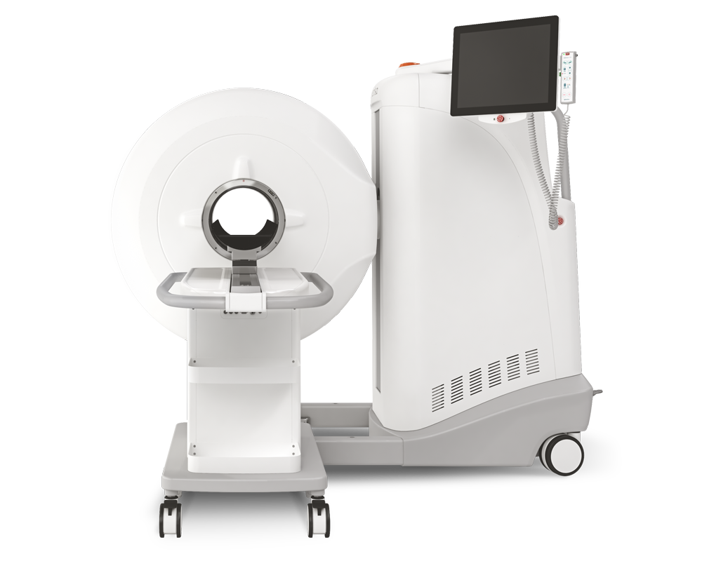
Results from MultiScan™ LFER PET/CT
- PET revealed an increased tracer uptake in the pulmonary lesions and mediastinal lymph nodes of all animals from the first scan obtained after infection and onwards
- the development of the lesions could be followed over time with PET and CT imaging as well
- [18F]DPA714 is a valuable radiotracer to visualize SARS-CoV-2-associated pulmonary inflammation, which coincided with activation of dendritic cells in blood

Fig. 4. Pulmonary lesion and inflammation process in a macaque after a SARS-CoV-2 infection visualized with [18F]DPA714. A. an example of a ground glass opacity (GGO) visualized with both CT and PET-CT. B. Transversal slices of the longitudinal follow-up of one animal (inflammation process is indicated with a white arrow).
Comment pouvons-nous vous aider?
N'hésitez pas à nous contacter pour obtenir des informations techniques ou à propos de nos produits et services.
Contactez-nous