MRI/PET multimodal imaging of the innate immune response in skeletal muscle and draining lymph node post vaccination in rats
2023.01.11.
Saaussan Madi,Fang Xie,[...],Shih-Hsun Cheng 1, Tolulope Aweda 1, Bhasker Radaram, Hasan Alsaid , Beat M Jucker
Frontiers in Immunology, 2023
Abstract
The goal of this study was to utilize a multimodal magnetic resonance imaging (MRI) and positron emission tomography (PET) imaging approach to assess the local innate immune response in skeletal muscle and draining lymph node following vaccination in rats using two different vaccine platforms (AS01 adjuvanted protein and lipid nanoparticle (LNP) encapsulated Self-Amplifying mRNA (SAM)). MRI and 18FDG PET imaging were performed temporally at baseline, 4, 24, 48, and 72 hr post Prime and Prime-Boost vaccination in hindlimb with Cytomegalovirus (CMV) gB and pentamer proteins formulated with AS01, LNP encapsulated CMV gB protein-encoding SAM (CMV SAM), AS01 or with LNP carrier controls. Both CMV AS01 and CMV SAM resulted in a rapid MRI and PET signal enhancement in hindlimb muscles and draining popliteal lymph node reflecting innate and possibly adaptive immune response. MRI signal enhancement and total 18FDG uptake observed in the hindlimb was greater in the CMV SAM vs CMV AS01 group (↑2.3 – 4.3-fold in AUC) and the MRI signal enhancement peak and duration were temporally shifted right in the CMV SAM group following both Prime and Prime-Boost administration. While cytokine profiles were similar among groups, there was good temporal correlation only between IL-6, IL-13, and MRI/PET endpoints. Imaging mass cytometry was performed on lymph node sections at 72 hr post Prime and Prime-Boost vaccination to characterize the innate and adaptive immune cell signatures. Cell proximity analysis indicated that each follicular dendritic cell interacted with more follicular B cells in the CMV AS01 than in the CMV SAM group, supporting the stronger humoral immune response observed in the CMV AS01 group. A strong correlation between lymph node MRI T2 value and nearest-neighbor analysis of follicular dendritic cell and follicular B cells was observed (r=0.808, P<0.01). These data suggest that spatiotemporal imaging data together with AI/ML approaches may help establish whether in vivo imaging biomarkers can predict local and systemic immune responses following vaccination.
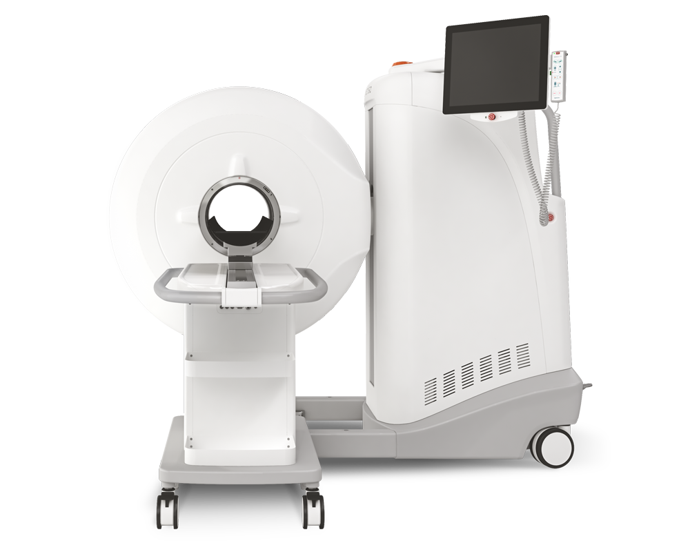
Results from MultiScan™ LFER PET/CT
- The present study represents the first application of a clinically translatable, combined MRI and PET imaging methodology for the simultaneous and non-invasive assessment of immune activation in muscle and draining lymph node following vaccination.
- the18FDG PET uptake in lymph node provided a unique measure of immune activity to differentiate vaccine platforms that could not be achieved using MRI

Fig 2, 18FDG PET images of rat hindlimb at 24 hr post Prime vaccine injection. Axial CT image with right popliteal lymph node identified for CMV SAM (A) and with 18FDG PET image coregistered with the CT image (B). Hindlimb 18FDG uptake in CMV SAM in Coronal (C) and Axial (D) planes. Coregistration of MRI and PET images (E). Orange arrows indicate popliteal lymph node, and yellow ROIs indicate gastrocnemius muscle region.
Comment pouvons-nous vous aider?
N'hésitez pas à nous contacter pour obtenir des informations techniques ou à propos de nos produits et services.
Contactez-nous