Metabolic Differences in Neuroimaging with [18F]FDG in Rats Under Isoflurane and Hypnorm–Dormicum
2025.01.03.
Aage Kristian Olsen Alstrup et al., Tomography 2025
Background: Anesthesia can significantly impact positron emission tomography (PET) neuroimaging in preclinical studies. Therefore, understanding these effects is crucial for accurate interpretation of the results. In this experiment, the authors investigated the effect of [18F]-labeled glucose analog fluorodeoxyglucose ([18F]FDG) uptake in the brains of rats anesthetized with two commonly used anesthetics for rodents: isoflurane, an inhalation anesthetic, and Hypnorm–Dormicum, a combination injection anesthetic.
Materials and Methods: Female adult Sprague Dawley rats were randomly assigned to one of two anesthesia groups: isoflurane or Hypnorm–Dormicum. The rats were submitted to dynamic [18F]FDG PET scan. The whole brain [18F]FDG standard uptake value (SUV) and the brain voxel-based analysis were performed.
Results: The dynamic [18F]FDG data revealed that the brain SUV was 38% lower in the isoflurane group after 40 min of image (2.085 ± 0.3563 vs. 3.369 ± 0.5577, p = 0.0008). In voxel-based analysis between groups, the maps collaborate with SUV data, revealing a reduction in [18F]FDG uptake in the isoflurane group, primarily in the cortical regions, with additional small increases observed in the midbrain and cerebellum.
Discussion and Conclusions: The observed differences in [18F]FDG uptake in the brain may be attributed to variations in metabolic activity. These results underscore the necessity for careful consideration of anesthetic choice and its impact on neuroimaging outcomes in future research.
Results from nanoScan® PET/MRI
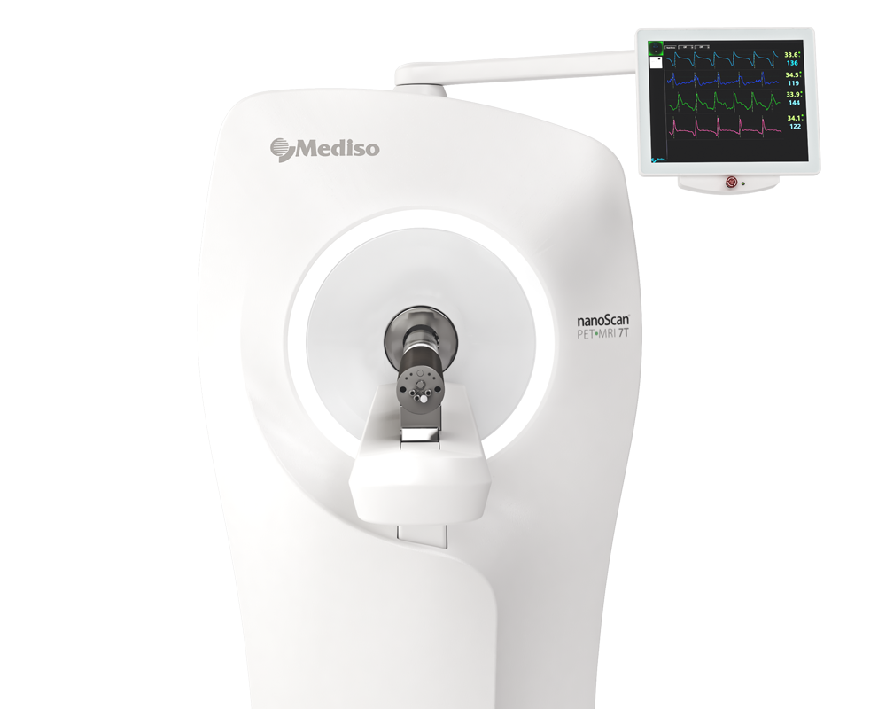
The twelve rats included in the study were placed in the Mediso nanoScan® PET/MRI 1T scanner one-by-one and briefly scanned in the MRI (for attenuation correction), then imaged using the PET scanner for 60 min after administration of [18F]FDG (18.7–35.2 MBq in 0.2–0.3 mL) intravenously in the tail catheter. Pulse, respiration frequency, and body temperature were monitored by using Mediso MultiCell™ system. Emission sinograms were iteratively reconstructed into a 23-frames (8 × 15, 4 × 30, 2 × 60, 2 × 120, 4 × 300, 3 × 600 s) and one frame of 20 min in the last part of the acquisition (Tera-Tomo™ 3D, 4 iteration and 6 subsets, as reconstruction method) after being submitted to MRI attenuation correction, normalized, and corrected for scatter and radioactivity decay. PET image analysis was performed with PMOD software (PMOD™ Technologies Ltd., Zurich, Switzerland, version 4.0). [18F]FDG PET images were registered to MRI template, and whole brain volume of interest (VOI) was applied for brain analysis. Standardized uptake values (SUV) were calculated and SUV average images were normalized for whole brain uptake to generate SUVR images. To assess the effects of anesthesia on other organs the heart uptake was evaluated as well, included in the same field of view (FOV) as the brain. For this purpose, analysis was performed using the PMODTM View tool, employing the automated hot 3D VOI tool to delineate the VOI for each animal’s heart. The data for the SUV curve and the average SUV for the 40–60 min interval, handled as static images, were included in the analysis.
Results showed the followings:
- By using the MultiCell™ system for vital signals monitoring it could be observed that there were no statistical differences in pulse, respiration rate, and body temperature between the two groups.
- The dynamic PET data revealed that after 25 min, the SUV for isoflurane group was lower than for Hypnorm–Dormicum data. One frame PET data (20 min of image acquisition between 40 and 60 min after [18F]FDG injection) showed an SUV 38% lower in the animals anesthetized with isoflurane after 40 min of acquisition.
- For the heart analysis, the dynamic PET data revealed higher uptake of [18F]FDG in the animals anesthetized with isoflurane after 9 min (Figure 3A). For the static image analysis of the last minutes of the image, 45% higher SUV could be observed in the isoflurane group when compared to Hypnorm–Dormicum animals (Figure 3B,D). The correlation between the brain and heart SUVs was not significant, but an inverse profile between the uptake in the heart and in the brain was showed; higher heart uptake and lower brain uptake in isoflurane animals and the opposite effect in the Hypnorm–Dormicum animals (Figure 3C).

Figure 3. Data representation for heart [18F]FDG analysis. The heart SUV curve for isoflurane (in red) and Hypnorm–Dormicum (in blue) groups (A). SUV data for the static image from 40 to 60 min after [18F]FDG administration (B). Data correlation for heart and brain SUV (C). Illustrative image of PET/MRI [18F]FDG uptake in SUV, the white arrows indicate the heart uptake. Note that there is a higher uptake in isoflurane animals when compared to Hypnorm–Dormicum animal (D). * p < 0.05; ** p < 0.01; *** p < 0.001.
Conclusion: Based on both prior research and the current findings, maintaining animals awake after [18F]FDG administration appears preferable for preserving neuronal function, especially in cortical regions, and for approximating biological conditions more closely. However, if dynamic imaging is necessary, careful consideration of study design, including the choice of anesthesia, is essential. Selecting the appropriate [18F]FDG uptake condition (anesthesia vs. awake) is critical in small animal [18F]FDG-PET studies, as altered baseline brain glucose metabolism levels could influence the biological effects researchers aim to observe.For future studies, it should be considered to include standardized fasting, measurements of plasma glucose before and after scanning, measurement of cerebral blood flow, and supplement the PET scans with ex vivo analysis.
Full article on mdpi.com
Comment pouvons-nous vous aider?
N'hésitez pas à nous contacter pour obtenir des informations techniques ou à propos de nos produits et services.
Contactez-nous