Investigation of Radiolabeled KISS1R Ligands as Promising Tools for Diagnosis and Treatment of Triple-Negative Breast Cancer
2026.03.03.
Harun Taş et al., Molecular Pharmaceutics, 2026
Summary
Kisspeptins (KPs) and their receptor KISS1R contribute to tumor progression and metastasis in cancers such as triple-negative breast cancer (TNBC), making KISS1R an attractive target for molecular imaging and targeted radionuclide therapy. However, early approaches using Ga-68/Lu-177–labeled KP-10 and KP-54 were limited by rapid proteolytic degradation and low tumor uptake. In this study, N-terminally functionalized derivatives of KP-10, KP-54, and the hybrid peptide KiSS-34 were synthesized and conjugated to DOTA and AF-488 while retaining high biological activity (EC50: 0.05–0.85 nM). Conventional antibody-based detection failed to reliably visualize KISS1R, whereas live-cell imaging with fluorescent KP analogues revealed rapid receptor internalization. Radiolabeled DOTA conjugates were obtained in high yields (≥95%), with [177Lu]Lu-DOTA-KiSS-34 showing the highest cellular uptake (4.8%) and internalization rate (45.9%). Importantly, PET/CT imaging, urine analysis, and in vitro studies demonstrated superior pharmacokinetics and in vivo stability for Ga-68/Lu-177-DOTA-KiSS-34 compared with KP-10 and KP-54 analogues, identifying it as a promising lead for KISS1R-targeted PET/CT imaging and radiotheranostics.
PET/CT imaging studies were conducted in normal, healthy mice. The mice were anesthetized with isoflurane and were retro-orbitally injected with 100–150 μL of radiotracers corresponding to 4–6 MBq of radiotracer per animal. Mice were positioned prone in the Mediso nanoScan® PET/CT small animal imaging system (Mediso Medical Imaging Systems, Budapest, Hungary), and static imaging was initiated 30 and 90 min post injection. A single PET field of view of 98.5 mm was imaged for 10 min, followed by a whole-body helical CT scan (50 kVp/980 μA and 720 projections). The images were reconstructed using Mediso Tera-TomoTM 3D PET iterative reconstruction (Mediso Medical Imaging Systems, Budapest, Hungary). Visualization and processing of the images were performed using a Mediso InterViewTM FUSION (Mediso Medical Imaging Systems, Budapest, Hungary). The scans were normalized to the injected activity and animal weight.
-
The PET/CT images revealed clearly different tissue distribution patterns among the three tracers (see Fig 1). The tracer [68Ga]Ga-DOTA-KP-54 exhibited strong and prolonged retention in the kidneys, indicating slow renal clearance and accumulation in the renal system. This behavior suggested less favorable pharmacokinetics for imaging applications because prolonged kidney retention can increase radiation dose and background signal.
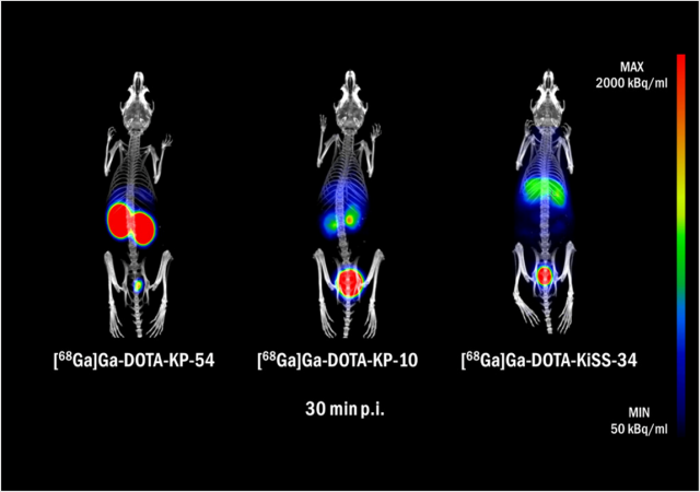
Fig 1. PET/CT images of healthy BALB/c mice injected with [68Ga]Ga-DOTA-KP-54, [68Ga]Ga-DOTA-KP-10, and [68Ga]Ga-DOTA-KiSS-34. Images are presented as maximum intensity projections of fused PET and CT at 30 min p.i.
-
When the peptide length was reduced to form [68Ga]Ga-DOTA-KP-10, the kidney retention was significantly decreased, and faster renal clearance was observed. This result demonstrated that structural shortening of the peptide improved pharmacokinetic behavior compared with the longer KP-54 analogue.
-
The tracer [68Ga]Ga-DOTA-KiSS-34, which consists of only six amino acids, showed similarly rapid renal clearance to KP-10 but with additional distinct biodistribution features. In particular, partial liver accumulation was observed with [68Ga]Ga-DOTA-KiSS-34, whereas this uptake was less pronounced for KP-54 and KP-10. The authors attributed this liver uptake to the presence of lipophilic amino acid residues such as AMBA and 2-Nal in the KiSS-34 structure.
-
An important PET-relevant result was obtained from the in vivo stability analysis of the tracers. Urine samples collected at 30 and 90 minutes after injection showed that [68Ga]Ga-DOTA-KP-54 and [68Ga]Ga-DOTA-KP-10 underwent significant degradation in vivo, as evidenced by altered chromatographic retention times. This degradation indicated that part of the PET signal from these tracers could originate from breakdown products rather than intact radiotracer molecules.
-
In contrast, [68Ga]Ga-DOTA-KiSS-34 remained stable in vivo and showed unchanged retention times during urine analysis. This stability demonstrated that the tracer maintained its structural integrity after administration and supported more reliable PET signal interpretation. The improved stability also suggested that radionuclide release due to degradation or decomplexation was unlikely for this tracer.
-
Overall, the PET/CT imaging data showed that [68Ga]Ga-DOTA-KiSS-34 had substantially lower kidney retention compared with KP-10 and KP-54 while maintaining favorable clearance characteristics. However, the increased liver accumulation observed with KiSS-34 indicated that further structural optimization might be required to reduce hepatic uptake and improve tumor-to-background contrast in future applications.


W czym możemy pomóc?
Skontaktuj się z nami aby uzyskać informacje techniczne i / lub wsparcie dotyczące naszych produktów i usług.
Napisz do nas