PD-1 blockade exacerbates Mycobacterium tuberculosis infection in rhesus macaques
2021.01.15.
Keith D Kauffman et al., Science Immunology, 2021
Summary
PD-1 (programmed death-1) is a coinhibitory receptor primarily expressed on activated CD4 and CD8 T cells that has been shown to limit the function of pathogen-specific T cells during chronic infection. The reactive expression of PD-L1 (PD-1 receptor ligand) on cancer cells turns off the T cells that are trying to attack the tumor. Therefore, blockade of PD-1 receptor or its ligands with monoclonal antibodies (mAbs) has become an attractive target in cancer therapy. Recognition of this pathway has led to suggestions that anti–PD-1 therapy might also boost T cell immunity in chronic infections including tuberculosis.
In this recent Science Immunology publication, Kauffman et al. examined the role of PD-1 during Mycobacterium tuberculosis (Mtb) infection of rhesus macaques. It was shown that the PD-1 blockade increased the number and functionality of Mtb-specific CD8 T cells, but not CD4 cells and was associated with increases in proinflammatory cytokines. However, animals treated with anti–PD-1 monoclonal antibody developed worse disease and higher granuloma bacterial loads compared with isotype control–treated monkeys.
These data indicate that negative regulation of immune responses is a critical aspect of host resistance to Mtb infection. Also, these findings suggest that the anti-PD-1 cancer therapy needs to be used cautiously in patients with a history of Mtb exposure.
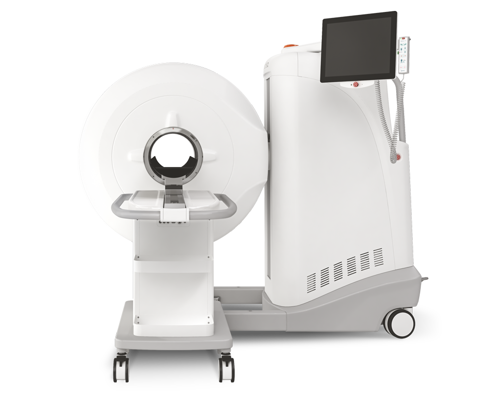
Results from MultiScan™ LFER
Mediso MultiScan™ Large Field of view Extreme Resolution (LFER) PET/CT system was used to follow the course of Mycobacterium tuberculosis (Mtb) infection in rhesus macaques. The animals were imaged before infection and every 2 weeks after infection beginning at 4 weeks for a maximum of eight PET-CT scans. [18F] FDG was injected intravenously (1 mCi/kg), and after 60 minutes incubation time a 20-min PET scan was acquired.

(C) Example PET-CT image from isotype control– (top) or PD-1 (bottom)–treated animals. (D) Fold change over week 4 value of total lung standardized uptake value (SUV) in isotype control (left) and PD-1–treated (right) animals.
Read the full articel on Science Immunology
How can we help you?
Don't hesitate to contact us for technical information or to find out more about our products and services.
Get in touch