Evaluation of the Therapeutical Effect of Matricaria Chamomilla Extract vs. Galantamine on Animal Model Memory and Behavior Using 18F-FDG PET/MRI
2024.05.09.
Roxana Iacob et al, Curr. Issues Mol. Biol., 2024
Summary
Alzheimer’s disease (AD) describes 60–70% of cases affecting more than 55 million people worldwide, a debilitating disease whose incidence could potentially triple in the next 30 years. The adult process of neurogenesis is altered in the earliest stages of Alzheimer’s disease (AD), by progressive and irreversible loss of memory, of higher cerebral functions and later, of the self-help skill. Structurally, the hippocampal areas show some rates of atrophy in the earliest stages of the disease, while the precuneus and posterior cingulate show consistent and increasing rates of atrophy later. These are useful indices of progression of AD.
The brain has the highest avidity for glucose, consuming about 20% of the energy derived from its metabolism. But in AD, glucose metabolism is dramatically decreased, resulting in synaptic dysfunction and neuronal death and thus the thinning of important areas of the cerebral cortex. The most involved hypometabolic cerebral regions are the hippocampus, posterior cingulate, precuneal, retrosplenial and limbic cortices.
The effectiveness of a long-term classic treatment of AD, represented by alkaloids with acetylcholinesterase-inhibiting proprieties (such as galantamine), induces a significant metabolic increase in the specific brain areas but does not determine a definitive cure of the disease. Also, it is already known that Scopolamine (Sco) induces cognitive impairment in experimental animals in all the cerebral regions of interest except the cerebellum and thalamus. These effects disappear with the co-administration of acetylcholinesterase inhibitors and Sco.
That is why there are all kinds of attempts to identify better treatments for the disease, including the Cioanca O. preclinical studies of antioxidant molecules that enter the brain and exert protective properties. As such, Matricaria chamomilla L. is a medicinal herb traditionally known to have anti-inflammatory, antimicrobial, antiviral, anxiolytic, and antidepressant proprieties. The hydroalcoholic extract of Matricaria chamomilla L (MCE) could be a potent neuropharmacological agent against amnesia via modulating cholinergic activity, neuroinflammation, and promoting antioxidant action in the rat hippocampus.
The memory-enhancing activity of Matricaria chamomilla hydroalcoholic extract (MCE) is already being investigated by behavioral and biochemical assays in scopolamine-induced amnesia rat models, while the effects of scopolamine (Sco) on cerebral glucose metabolism are examined as well. Nevertheless, the study of the metabolic profile determined by an enriched MCE has not been performed before. The present experiments compared metabolic quantification in characteristic cerebral regions and behavioral characteristics for normal, only diseased, diseased, and MCE- vs. Galantamine (Gal)-treated Wistar rats. A memory deficit was induced by Sco injections. The memory assessment comprised three maze tests. Glucose metabolism was quantified after the 18F-FDG PET examination.
The right amygdala, piriform, and entorhinal cortex showed the highest differential radiopharmaceutical uptake of the 50 regions analyzed. Rats treated with MCE show metabolic similarity with normal rats, while the Gal-treated group shows features closer to the diseased group. Behavioral assessments videnced a less anxious status and a better locomotor activity manifested by the MCE-treated group compared to the Gal-treated group.
Results from nanoScan PET/MRI
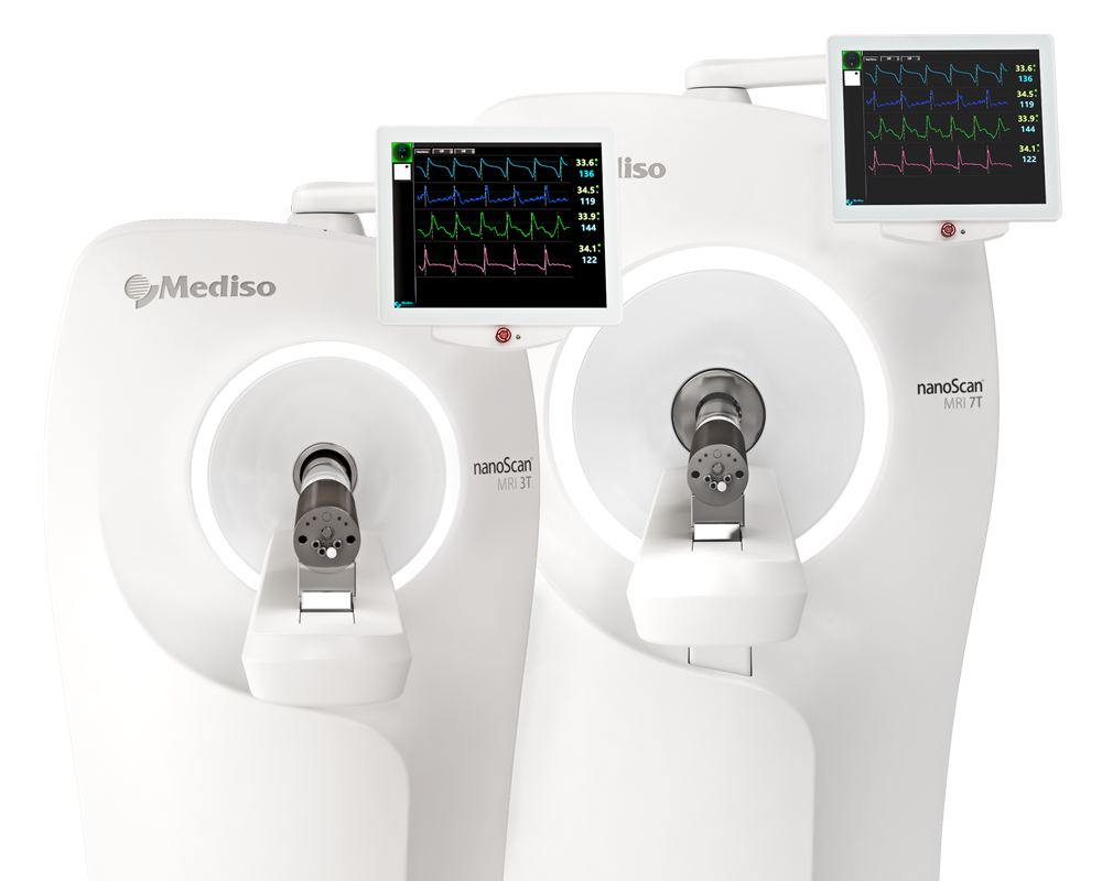
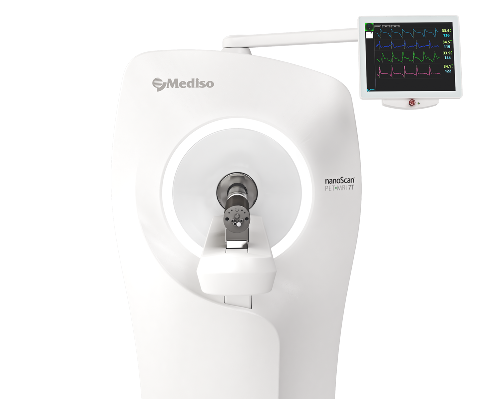
The 18F-FDG PET scans were obtained using an animal-dedicated PET/MRI scanner (nanoScan, Mediso Ltd., Budapest, Hungary). 3D PET images were reconstructed using a Maximum Likelihood Estimation Method (MLEM) algorithm (8 iterations and 3 subsets), with a slice thickness of 3 mm. The acquisitions were centered on the head region of each rat and were accomplished in the third week of the experiment. Image acquisitions were performed in the prone position, with the PET scan 45 min after the radiotracer administration. MRI data acquisition comprised three series of images as follows: a Fast Scout Sagittal Series, a GRE 3D Multi FOV Series, and a multi-echo 2D fast spin echo sequence (T2 FSE 2D).
PET data acquisition comprised an image series, including a static acquisition of 30 min Tera-Tomo 3D Full Model, with a MLEM of image reconstruction (8 iterations and 3 subsets), 400–600 keV, 1:5, slice thickness: 3 mm.
In all subjects, 18F-FDG uptake was visible in all cerebral areas and the cerebellum. Furthermore, the quantitative 18F-FDG accumulation expressed in the SUVmax, SUVmean, and SUVglc values was calculated for AD characteristic regions of the brain, including the whole brain, cingulate cortex, precuneus, caudate putamen, callosus cortex, entorhinal cortex, retrosplenial, globus pallidus, accumbens, amygdala, hippocampus, thalamus, and hypothalamus.

Figure 1. Coronal section of: (a) 18F-FDG PET brain template, on a Rainbow Color Scale; (b) reference MRI template, on a Gray Color Scale; and (c) fused 18F-FDG PET/MRI. Letter “R” represents the right side of the image.

Figure 2. Coronal section of the (a) Schwartz atlas VOI template, on a Gray Color Scale; (b) 18F-FDG PETWistar brain rat, on a Rainbow Color Scale; and (c) fused images highlight the automated VOI for SUVglc parametric images analysis.
The PC (positive control) rats showed significantly higher Whole Brain SUVglc levels (2.49 ± 0.76 g/mL, range 0.63–8.09 g/mL) than the NC (negative control) rats (1.96 ± 0.49 g/mL, range 0.61–5.38 g/mL). The right amygdala showed the greatest increase (37% difference) in Sco-injected rats, while the right raphe nucleus showed the least (9% difference). The left (6% difference) and right (4% difference) pons showed a significant decrease in SUVglc levels. After 1 and 2 weeks of treatment, the Sco-induced rats treated with Matricaria chamomilla extract show metabolic similarity with normal rats, in the AD-specific cerebral regions, upon 18F-FDG PET examination. The Gal-treated group shows features closer to the PC group, as can be seen in Figure 3.

Figure 3. Metabolic effects of Matricaria chamomilla extract in AD specific cerebral regions, by 18F-FDG PET examination, after chronic treatment of Sco-induced cognitively impaired rats.
These metabolic features are maintained regardless of the SUV calculation method (the maximum uptake, the mean uptake, or the glucose corrected formula) for glucose metabolism in all the characteristic cerebral regions. Sco-induced and Chamomilla-treated rats present SUV values closer to those of normal rats, as shown in Figure 4.

Figure 4. Comparison of metabolic features (SUVmax, SUVmean and SUVglc) of chronic Matricaria chamomilla extract treatment with Galantamine treatment, Positive and Negative Control groups, by 18F-FDG PET examination, in Sco-induced cognitively impaired rats.
Conclusion
- These findings prove evident metabolic ameliorative qualities of MCE over Gal classic treatment, suggesting that the extract could be a potent neuropharmacological agent against amnesia of AD origin.
- Rats treated with MCE show metabolic similarity with normal rats, while the Gal-treated group shows features closer to the diseased group.
- Behavioral assessments evidenced a less anxious status and a better locomotor activity manifested by the MCE-treated group compared to the Gal-treated group.
- In AD treatments, because of the ease of administration, these hydroalcoholic extract could represent a definite area of research in the future.
- This study proved better performances for the Matricariatreated group compared to the Gal-treated group in all the behavioral evaluations (short-term memory, spontaneous and induced activity, and anxietymanagement), aswell as the metabolic ameliorative qualities ofMCE over the Gal classic treatment in AD-specific cerebral areas.
- This research will allow for further translational studies of this natural extract on human patients because it has minimal to no side effects thus increasing the compatibility of the treatment to patients.
Full article on mdpi.com
How can we help you?
Don't hesitate to contact us for technical information or to find out more about our products and services.
Get in touch
