Porphyrin-lipid nanotheranostics for multimodal imaging of nodal disease in preclinical oral cancers
2026.05.29.
Michael S. Valic et at., Theranostics, 2026
Summary
Cervical lymph node metastases in oral cancer patients are a frequent occurrence and important prognostic factor. Anatomical and molecular imaging modalities can identify neck metastases with varying sensitivity and specificity but perform poorly in clinically negative neck nodes with microscopic disease. In this article the authors investigated the use of porphyrin-lipid nanotheranostics (PS) for multimodal detection of neck disease in preclinical models of oral cancer. PS nanoparticles were radiolabelled with positron-emitting Copper-64 and uptake in the tumour and cervical lymph nodes was measured with serial PET/MR imaging.
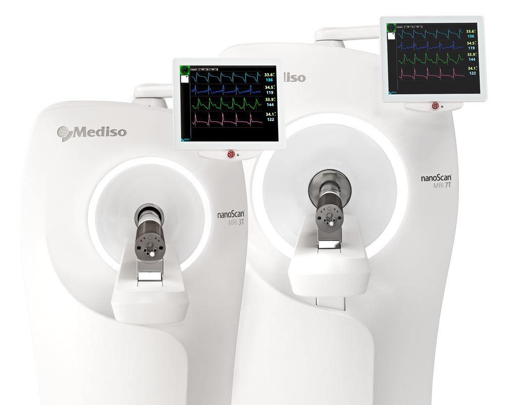
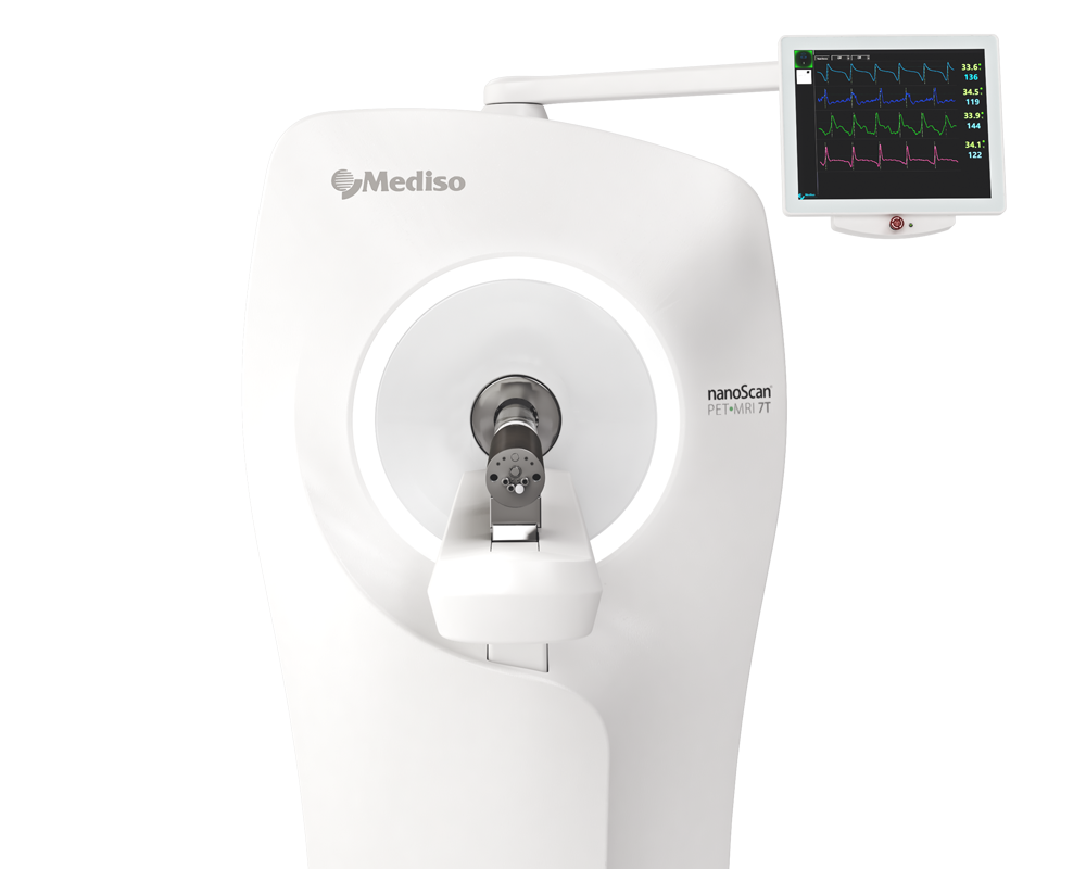
Results from nanoScan® PET/MRI
PET/MR imaging was performed using a 1T PET/MR small animal scanner (nanoScan® Mediso, Budapest, Hungary). For intratumoural lymphatic mapping experiments, 64Cu-PS (80–120 MBq 64Cu, 0.5 mg pyro-lipid, IT) were administered into the tongue tumour. PET/MR imaging was performed at timepoints: 1 h, 3 h, 6 h, 12 h, 24 h, 48 h, and 72 h post-IT injection. PET acquisition times ranged from 10–25 min and T1 3D material maps were collected for MRI-based attenuation correction using parameters: 256 x 182 matrix, 0.35 x 0.35 x 0.60 mm3 voxel size, 18.98 ms repetition time, 2.9 ms echo time, 15° flip angle.
For comparison experiments between systemically injected PET radiotracers, 18F-FDG was injected and imaged 24 h prior to experiments involving 64Cu-PS. 18F-FDG (46 MBq 18F/kg, IV) was administered via the tail vein and PET/MR imaging performed after ~45 min of uptake. PET acquisition time was 10 min using the same imaging parameters described above. The following day, 64Cu-PS (250–500 MBq 64Cu/kg, 0.5–1.0 mg/kg pyro-lipid, IV) were administered via tail vein and PET/MR imaging repeated as described above at 3 h and 24 h post-IV injection using a 15–20 min PET acquisition time. All PET images were reconstructed using parameters: 400-600 keV energy window, 105 x 105 x 237 matrix, 0.4 mm3 voxel size.
- Using the Mediso nanoScan® PET/MR scanner, serial PET/MR imaging was performed from 1 to 72 hours after intratumoral administration of 64Cu-PS. The PET images clearly visualized sentinel and downstream cervical lymph nodes as early as 1 hour after injection, with the highest contrast observed between 12 and 24 hours (see Figure 1.). Metastatic nodes tended to accumulate and retain more nanoparticle than benign nodes. However, despite excellent visualization of lymphatic drainage pathways, the PET signal differences between metastatic and benign nodes were not statistically significant. Consequently, intratumoral 64Cu-PS PET/MR was valuable for lymphatic mapping and surgical planning but was insufficient as a stand-alone diagnostic method for nodal staging.
Fig 1. Representative MIPs from serial 64Cu-PS PET/MR imaging post-IT injection (80–120 MBq 64Cu, 0.5 mg pyro-lipid). Pathological staging of nodes indicated on 3 h image. 64Cu PET signal intensity arbitrarily scaled to the 1 h timepoint. (B) Healthy rats (i.e., without tongue tumours) administered 64Cu-PS into the tongue and serially imaged. 64Cu PET signal intensity scaling as in (A).

- The authors then evaluated systemic administration of 64Cu-PS. PET/MR imaging performed on the scanner showed that at 3 hours post-injection most nanoparticles remained in the circulation, resulting in relatively high background activity. By 24 hours, background activity had decreased and the tongue tumors and cervical lymph nodes became clearly visible (Figure 2.).
The PET/MR images demonstrated strong passive accumulation of 64Cu-PS within the tumors, with tumor SUVmean increasing significantly between 3 and 24 hours. Comparison with 18F-FDG PET showed that the 24-hour 64Cu-PS PET images achieved tumor and nodal contrast comparable to conventional FDG imaging. Histological validation further confirmed that PET signal corresponded closely with nanoparticle fluorescence and autoradiographic localization within tumor tissue.
Fig 2. (A) Representative MIPs of 18F-FDG PET/MR images 45 min post-IV injection (46 MBq 18F/kg), and 64Cu-PS PET/MR images from 3 h and 24 h post-IV injection (250–500 MBq 64Cu/kg, 0.5–1.0 mg/kg pyro-lipid) in tumour-bearing rats. Magnified view of outlined neck nodes in insets. Arrows denote tumour. 64Cu PET signal intensity arbitrarily scaled to the 3 h timepoint. (B) Representative MIPs of 64Cu-PS PET/MR images from healthy rats (i.e., without tongue tumours).

- Figure 3. presents the main PET imaging results. At 24 hours after intravenous injection, the scans visualized 83% of harvested cervical lymph nodes, including most metastatic nodes. Quantitative PET analysis showed significantly higher 64Cu-PS uptake in metastatic lymph nodes compared with benign nodes. Importantly, 64Cu-PS PET outperformed conventional 18F-FDG PET. Whereas FDG uptake did not significantly differ between metastatic and benign nodes, 64Cu-PS produced a substantially better ROC performance (AUC ≈ 0.78), demonstrating superior ability to identify microscopic nodal disease.
Fig 3. Representative MIPs of 64Cu-PS PET/MR images from 24 h post-IV injection (250–500 MBq 64Cu/kg, 0.5–1.0 mg/kg pyro-lipid). 64Cu-PS uptake in pN0 vs pN+ staged nodes.

Overall, the study demonstrated that 64Cu-PS PET/MR imaging on the Mediso nanoScan platform provides a sensitive multimodal approach for detecting occult cervical lymph node metastases, outperforming conventional MRI morphology assessment and showing better diagnostic accuracy than 18F-FDG PET, while also enabling complementary fluorescence-guided surgical navigation.
Full article on thno.org
How can we help you?
Don't hesitate to contact us for technical information or to find out more about our products and services.
Get in touch