PET Evaluation of the Novel F‑18 Labeled Reversible Radioligand [18F]GEH200449 for Detection of Monoamine Oxidase‑B in the Non- Human Primate Brain
2023.08.16.
Katarina Varnäs et al., ACS Chem. Neurosci., 2023
Summary
The article focuses on the enzymes Monoamine Oxidase A and B (MAO-A, MAO-B), which play a crucial role in the metabolism of monoamine neurotransmitters in the brain. These enzymes are involved in the pathophysiology and treatment of various neurological and psychiatric disorders. Non-selective monoamine oxidase inhibitors possess antidepressant properties, while MAO-B selective inhibitors are widely used in the pharmacological treatment of Parkinson's disease. Furthermore, MAO-B is abundantly present in brain glial cells, and increased expression has been observed in reactive astrocytes. In Alzheimer's disease, the overexpression and elevated levels of MAO-B have been linked to the accumulation of amyloid β-peptides, responsible for the formation of amyloid brain deposits.
Imaging of MAO-B binding using positron emission tomography (PET) can be valuable for assessing drug effects, and it is increasingly applied as a potential biomarker for monitoring astrogliosis in neurodegenerative diseases. PET imaging of MAO-B utilizes radioligands labeled with enzyme inhibitors or metabolic trapping compounds. Several MAO-B PET radioligands have been developed, such as [11C]L-deprenyl-D2. However, [11C]L-deprenyl-D2 has limitations, including irreversible binding and the formation of radioactive metabolites that can enter the brain, necessitating the development of new radioligands. One example is [11C]SL25.1188, characterized as a reversible, selective MAO-B binding radioligand, and it has been used in studies of patients with depression and post-traumatic stress disorder.
To facilitate broader clinical research, MAO-B tracers are labeled with the longer-lived fluorine-18 isotope (with a half-life of 110 minutes, compared to 20.4 minutes for carbon-11). This includes [18F]SMBT-1, which has been employed in studies of Alzheimer's disease patients. Additionally, the MAO-B radioligand [18F]GEH200449 has been investigated, and promising results, such as specific binding and low non-specific binding in human brain tissue, have been found. In the current study, the suitability of [18F]GEH200449 as a PET radioligand was further assessed in non-human primates of the Macaca fascicularis.

Figure1.Time curves for [18F]GEH200449 brain regional radioactivity in NHP#4 at baseline (A) and after pretreatment with 0.5 mg/kg of L-deprenyl (B) or 0.75mg/kg of rasagiline (C) at 45 min before radioligand injection and after displacement with 0.5 mg/kg of L-deprenyl at 25 min after radioligand injection (D).Arrow indicates start of L-deprenyl injection. CAU; PUT; THA; PFC; CER; OC and SUV.
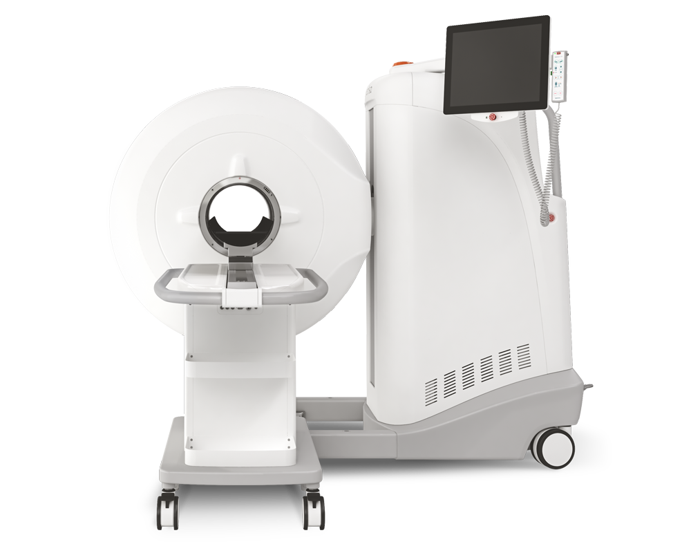
Results from MultiScan™ LFER150 PET/CT
The study involved 13 PET measurements in five NHPs. Due to technical challenges with arterial cannulation, arterial blood data could not be obtained for two PET measurements in NHP#2 and for the pretreatment studies with AZD9272, rasagiline (0.25mg/kg), and fenobam in NHPs#3−5.
The whole-brain radioactivity peaked rapidly at 3.4−5.2 SUV (4.0−6.4%ID) within 1.8−7.5 minutes after IV injection of [18F]GEH200449, making it suitable for regional binding analysis. PET images and regional radioactivity time curves showed high binding in the striatum and thalamus, with lower binding in the cortex and cerebellum, consistent with the known localization and levels of MAO-B.
-
[18F]-GEH200449 binding was reduced when pretreated with L-deprenyl or rasagiline before radioligand injection, indicating reversible binding to MAO-B.
-
AZD9272 and fenobam also reduced [18F]-GEH200449 binding, confirming binding to MAO-B, including its secondary binding site.
-
The fraction of unchanged [18F]-GEH200449 in plasma decreased rapidly after injection.
-
Kinetic models (1-TC and 2-TC) and the Logan graphical analysis were used for data interpretation.
-
The 2-TC model was preferred for most measurements, but VT values showed uncertainty in some regions and NHPs.
-
This uncertainty may be due to radiometabolites affecting the signal, requiring further investigation.
-
The Logan graphical analysis is recommended as the preferred method for quantitative analysis due to parameter estimate uncertainty.
Radioligand binding inhibition was observed in all brain structures in PET images (Figure 2) after drug administration. This indicates the absence of a suitable reference region for quantifying radioligand binding. The occupancy at MAO-B was calculated for pretreatment experiments with L-deprenyl and rasagiline. Using a graphical analysis of VT, the occupancy of L-deprenyl was 97% at the 0.5 mg/kg dose level and 82% at the 1.0 mg/kg dose level. For rasagiline at the 0.75 mg/kg dose level, the occupancy was 97% (Figure S4). Occupancy could not be calculated for 0.25 mg/kg rasagiline due to the unavailability of arterial plasma samples.

Figure2. Fused MR and PET images showing brain radioactivity after injection of [18F]GEH200449 in NHP#4 at baseline (A) and following administration of 0.5 mg/kg L-deprenyl (B),0.25mg/kg rasagiline (C),or 0.75mg/kg rasagiline (D).Average images for 123 min.SUV.

Figure3. (A) Time curve for radioactivity in putamen with model fits for the one-(1-TC) and two-tissue compartment (2-TC) models for NHP#3. (B) Total distribution volume (VT) obtained by the Logan graphical analysis plotted versus that obtained by the 2-TC model. VT was not included for the cerebellum in NHP#4, or for the occipital cortex in NHPs#4 and#5 since the2-TC model fits yielded implausible estimates of VT(>500mL cm−3) for these measurements and regions.
- Radiochemistry: The precursor (GEH200452) and non-radioactive reference standard (GEH200449) were supplied by GE Healthcare. [18F]GEH200449 was synthesized from GEH200452 as previously described. Its radiochemical purity was >99%, and the molar radioactivity ranged from 12 to 134 GBq/μmol, corresponding to an injected mass of 0.3 to 3.5 μg.
- Non-Human Primates (NHPs): This study was approved by the Animal Ethics Committee of the Swedish Animal Welfare Agency. Two male and three female NHPs (M. fascicularis, weighing 4.2-8.8 kg) were used. Anesthesia was induced with ketamine hydrochloride and maintained with ketamine and xylazine hydrochloride. Vital signs were monitored throughout the procedure.
-
PET Data Acquisition: PET measurements were conducted using Siemens Molecular Imaging's high-resolution research tomograph system for NHPs #1-4 and Mediso Ltd.'s LFER 150 PET/CT system for NHP #5. Each measurement was performed on a separate day, with at least a 6-week gap between experiments in the same NHP. Baseline PET measurements were initially done for all five NHPs. Subsequent drug inhibition binding studies were carried out using two selective MAO-B inhibitors, L-deprenyl and rasagiline, as well as two compounds, fenobam and AZD9272, known for their affinity toward MAO-B.
PET measurements included L-deprenyl administration in two NHPs, rasagiline in one NHP, fenobam in one NHP, and AZD9272 in one NHP. The test compounds were administered via intravenous infusion starting 45 minutes before the PET measurement for L-deprenyl and rasagiline, and 15 minutes before for fenobam and AZD9272.
To confirm [18F]GEH200449 binding reversibility, displacement PET measurements were conducted using L-deprenyl in two NHPs, administered 25 minutes after radioligand injection. Additional experimental details are available in Table S1.
During PET data acquisition, [18F]GEH200449 was injected as a bolus into a sural vein in a sterile phosphate buffer solution (pH = 7.4) at a radioactivity level of 107-167 MBq. Emission data were collected for 123 minutes, and arterial blood samples were taken at various intervals to determine the fraction of unchanged radioligand in plasma, as described in previous procedures.
-
PET data analysis: Dynamic images were reconstructed following previous methods. Regions of interest (ROIs) were manually drawn on T1-weighted MRI scans for various brain regions, including the whole brain, caudate nucleus, putamen, thalamus, occipital cortex, prefrontal cortex, and cerebellum. These ROIs were then applied to PET images after coregistration with the MRIs using PMOD software.
Time-activity curves for [18F]GEH200449 binding in different brain regions at baseline were analyzed using kinetic modeling, including 1-TC and 2-TC models, to calculate VT values. Model selection was based on Akaike information criterion and F statistics. Regional VT values were also determined using the Logan linear graphical method with a fixed time point (t*) of 40 minutes.
To estimate occupancy at [18F]GEH200449 binding sites, changes in regional VT values before and after drug administration were analyzed following a graphical procedure outlined in the literature. These analyses were conducted using PMOD software.
Conclusion
The PET evaluation of the novel MAO-B radioligand [18F]GEH200449 in NHPs demonstrated its high brain exposure and a regional brain distribution consistent with the known localization of MAO-B. The binding of [18F]GEH200449 could be effectively inhibited by the administration of reference MAO-B ligands before or after radioligand administration. The Logan graphical analysis method was chosen as the preferred approach for quantifying [18F]GEH200449 binding. These findings strongly support the suitability of [18F]GEH200449 as a reversible MAO-B radioligand for future human studies.
Full article on pubs.acs.org
W czym możemy pomóc?
Skontaktuj się z nami aby uzyskać informacje techniczne i / lub wsparcie dotyczące naszych produktów i usług.
Napisz do nas