Isoniazid and rifapentine treatment effectively reduces persistent M. tuberculosis infection in macaque lungs
2022.11.03.
Riti Sharan et al, The Journal of Clinical Investigation, 2022
Abstract
A once-weekly oral dose of isoniazid and rifapentine for 3 months (3HP) is recommended by the CDC for treatment of latent tuberculosis infection (LTBI). The aim of this study is to assess 3HP-mediated clearance of M. tuberculosis bacteria in macaques with asymptomatic LTBI. Twelve Indian-origin rhesus macaques were infected with a low dose (~10 CFU) of M. tuberculosis CDC1551 via aerosol. Six animals were treated with 3HP and 6 were left untreated. The animals were imaged via PET/CT at frequent intervals. Upon treatment completion, all animals except 1 were coinfected with SIV to assess reactivation of LTBI to active tuberculosis (ATB). Four of 6 treated macaques showed no evidence of persistent bacilli or extrapulmonary spread until the study end point. PET/CT demonstrated the presence of significantly more granulomas in untreated animals relative to the treated group. The untreated animals harbored persistent bacilli and demonstrated tuberculosis (TB) reactivation following SIV coinfection, while none of the treated animals reactivated to ATB. 3HP treatment effectively reduced persistent infection with M. tuberculosis and prevented reactivation of TB in latently infected macaques.
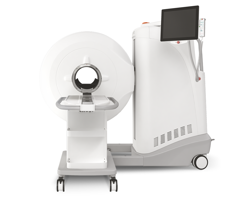
Results from MultiScan™ LFER PET/CT
- Coinfection with SIV led to TB reactivation in untreated animals, as demonstrated by the presence of numerous granulomatous lesions detected by CT scans
- PET scans demonstrating gradual progression in TB pathology from week 26 after TB infection up to necropsy with multiple new lung lesions
- PET scans of 6 animals treated with 3HP demonstrating no new lung lesions

CT scans of (A) control and (B) 3HP-treated rhesus macaques at weeks 8–10, 22, and 26 after TB infection and at study end point. Animal 31438 was an active progressor and was not administered SIV. In the longitudinal CT scans performed, macaques in the 3HP treatment group reported resolving lung lesions as early as 2–4 weeks after 3HP treatment initiation (black arrows), while there were no new lung lesions, and preexisting lung lesions resolved further at 10 weeks after 3HP initiation (black arrows)

(A) PET scans of 6 untreated control animals demonstrating gradual progression in TB pathology from week 26 after TB infection up to necropsy with multiple new lung lesions, and increased size of previously reported nodular lung lesions. (B) PET scans of 6 animals treated with 3HP demonstrating no new lung lesions, (C) granuloma counts, (D) lung lesion volume, (E) lung lesion activity, (F) lung SUVmax, and (G) total lung activity at weeks 26 and necropsy in treated and control animals. Data are represented as mean ± SEM. Significance was determined using 2-way ANOVA or multiple 2-tailed t tests using Holm-Šidák method, *P < 0.05; **P <0.01; ***P < 0.001.
Publication link: https://www.jci.org/articles/view/161564
W czym możemy pomóc?
Skontaktuj się z nami aby uzyskać informacje techniczne i / lub wsparcie dotyczące naszych produktów i usług.
Napisz do nas