Quantitative validation of Monte Carlo SPECT simulation: application to a Mediso AnyScan GATE simulation
2023.09.30.
Sophia et al., EJNMMI Physics, 2023
Background
Monte Carlo (MC) simulations are used in nuclear medicine imaging as they provide unparalleled insight into processes that are not directly experimentally measurable, such as scatter and attenuation in an acquisition. Whilst MC is often used to provide a ‘ground-truth’, this is only the case if the simulation is fully validated against experimental data. This work presents a quantitative validation for a MC simulation of a single-photon emission computed tomography (SPECT) system.
Methods
An MC simulation model of the Mediso AnyScan® TRIO SPECT/CT/PET system installed at the UK National Physical Laboratory was developed in the GATE (Geant4 Application for Tomographic Emission) toolkit. Components of the detector head and two collimator configurations were modelled according to technical specifications and physical measurements. Experimental detection efficiency measurements were collected for a range of energies, permitting an energy-dependent intrinsic camera efficiency correction function to be determined and applied to the simulation on an event-by-event basis. Experimental data were collected in a range of geometries with 99mTc for comparison to simulation. The procedure was then repeated with 177Lu to determine how the validation extended to another isotope and set of collimators.
Phantom acquisitions
Experimental acquisitions of 99mTc and 177Lu were performed with LEHR and MLEGP collimators, respectively. Images of each isotope were acquired with a uniform and a localised distribution of activity. A cylinder with 20 cm internal diameter and height was filled with a uniform solution of 99mTc. A commercial cylindrical Jaszczak phantom was used for the 177Lu acquisition; with an internal height of 18.6 cm and a diameter of 21.6 cm. A commercial NEMA phantom with six active spheres in an inactive background of water was used for both isotopes. The same activity concentration was dispensed to all spheres. Water was used as a scattering medium due to its similar density to human tissue. All phantom scans were performed in dual-head acquisition mode with a fixed detector radius of 250 mm. One emission (EM) and one scatter (SC) windows were simultaneously acquired for 99mTc imaging. A larger (5%) scatter window was used for the cylindrical phantom. 20% emission and 6% scatter windows were acquired for the two emissions of 177Lu.
Simulation model
A full MC simulation model was created for the Mediso AnyScan SPECT system based on specifications provided by the manufacturer and physical measurements. GATE version 8.2 with Geant4 version 10.05 was used. A detector head model was created and positioned for either dual or triple head configuration, as per the physical camera. Each head contained a 9.5 mm NaI thick crystal set as a ‘sensitive volume’, meaning all interactions within the volume were recorded. Geometric structures for the glass light-guide and the compartment housing the signal electronics were modelled in the simulation, as they were found to contribute to scatter into the crystal (as seen in other work ). The individual photomultiplier tubes were not defined in the simulation; instead, a general ‘back-compartment’ material was used to mimic the density of the electrical components, as suggested in. In addition to SPECT, the AnyScan SCP system has a computed tomography (CT) and a positron emission tomography (PET) ring, but these were not included in the simulation.
Models for LEHR and MLEGP collimators were defined as uniform blocks of lead with a rectangular array of hexagonal holes according to technical specifications of hole size and septal thickness. An aluminium touch-plate was included in front of the collimator and the detector head was surrounded by lead shielding. The plastic casing surrounding the heads was not included in the simulation. A visualisation of the simulation model with a geometric cylindrical phantom on the patient bed.
The patient bed was defined from a CT image, in order to accurately describe its internal structure.
The CT scan of the phantoms was used to define the source geometry and attenuation distribution in the simulation. The Hounsfield unit (HU) value of each voxel was attributed to a material density and elemental composition. The CT images were manually segmented to determine regions for activity. The CT scan was used to ensure accurate replication of the positioning and material composition of the phantom in the simulation. The ‘ImageNestedParameterisedVolume’ feature in GATE was used to speed-up the simulation of voxelised volumes. For the calibration ampoules and point sources, geometric source and phantom distribution were used due to the more simple geometry and material composition.

Top: A photograph of the SPECT system in dual head configuration. The third head (left) is not used to acquire data in this mode. Bottom: A visualisation of the simulation geometry with key components labelled. Each detector head is surrounded by lead shielding; this is shown as a wire frame here but fully encases each head.

Top: experimental and simulated projection images for the NEMA phantom of 99mTc for projection slice 38 is shown. The images have been cropped to 100x100 pixels. Bottom: the vertically averaged counts along a rectangular profile (shown in white on the inset) of the simulated and experimental image. The percentage residuals are shown with the ±3σ uncertainty of the sensitivity ratio shown as dashed lines
Results
The simulation’s spatial resolution, sensitivity, energy spectra and the projection images were compared with experimental measurements. The simulation and experimental uncertainties were determined and propagated to all calculations, permitting the quantitative agreement between simulated and experimental SPECT acquisitions to be determined. Statistical agreement was seen in sinograms and projection images of both 99mTc and 177Lu data. Average simulated and experimental sensitivity ratios of (0.991±0.0110) were seen for emission and scatter windows of 99mTc, and (0.897±0.0140) and (0.839±0.0140) for the 113 and 208 keV emissions of 177Lu, respectively.
Conclusion
It is essential to determine the validity of any simulation before it can be used to generate data. In this work, a Monte Carlo simulation of the Mediso AnyScan SCP system was created in the GATE simulation package and used to assess a quantitative validation methodology. The energy-dependent intrinsic efficiency was measured experimentally and used to create an efficiency-correction function which was applied to the simulation. The corrected simulation was then compared to several experimental observables to validate the simulation and quantify its agreement with the physical camera. A simulation will never perfectly replicate a physical system, particularly in nuclear medicine applications where full simulation of the photon-detection chain is generally too computationally expensive. This study was performed to quantify the similarity between the simulation and the physical camera. Energy spectra, planar and tomographic images were compared. Statistical agreement was seen in simulated and experiential images of both 99mTc and 177Lu. A quantitative validation has now been established, so the simulation can be used with confidence for other applications such as energy window optimisation, scatter estimation and acquisition parameter testing. Furthermore, this quantitative validation methodology can be applied to other SPECT systems and MC packages.
Full article on EJNMMI Physics.
-
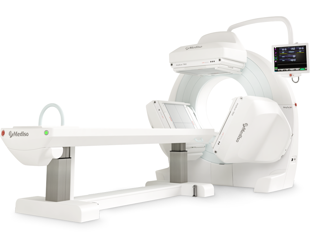
AnyScan® TRIO SPECT
Ultra-fast Triple-NaI-Detector SPECT for wide range of clinical applications
-
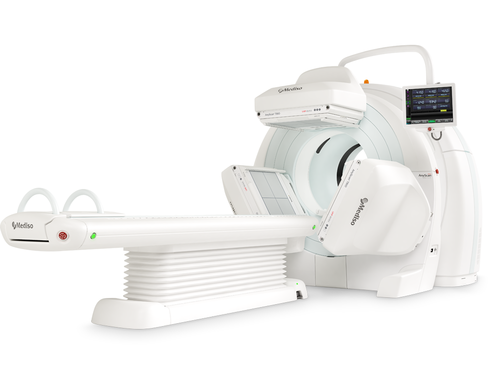
AnyScan® TRIO SPECT/CT
Ultra-fast Triple-NaI-Detector SPECT system for wide range of clinical applicati...
-
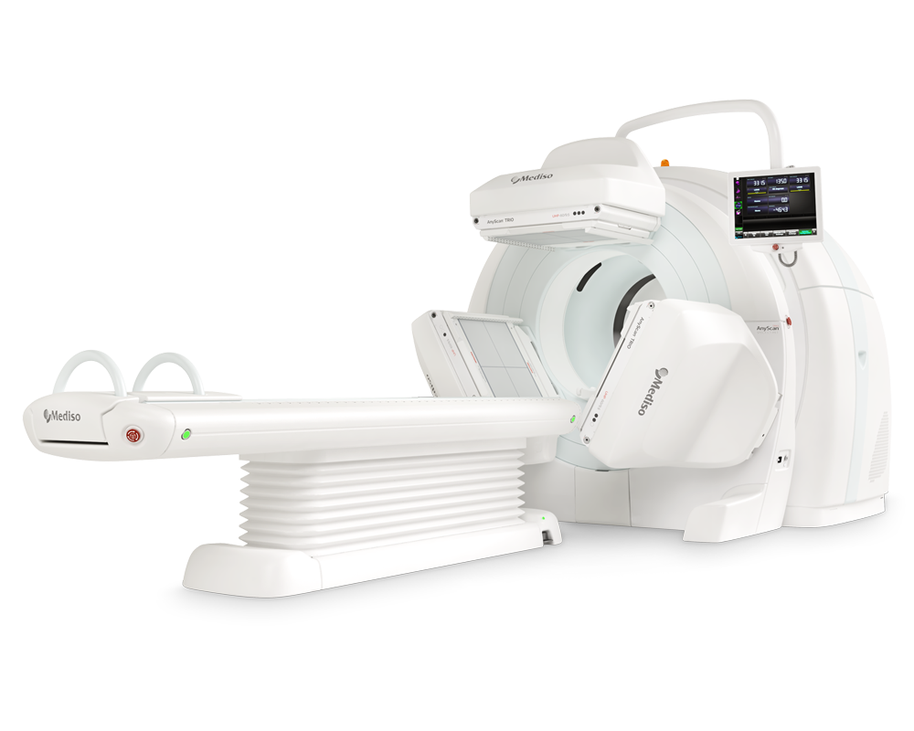
AnyScan® TRIO SPECT/CT/PET
An integrated triple-detector SPECT/CT and PET/CT system in a single-room instal...
-
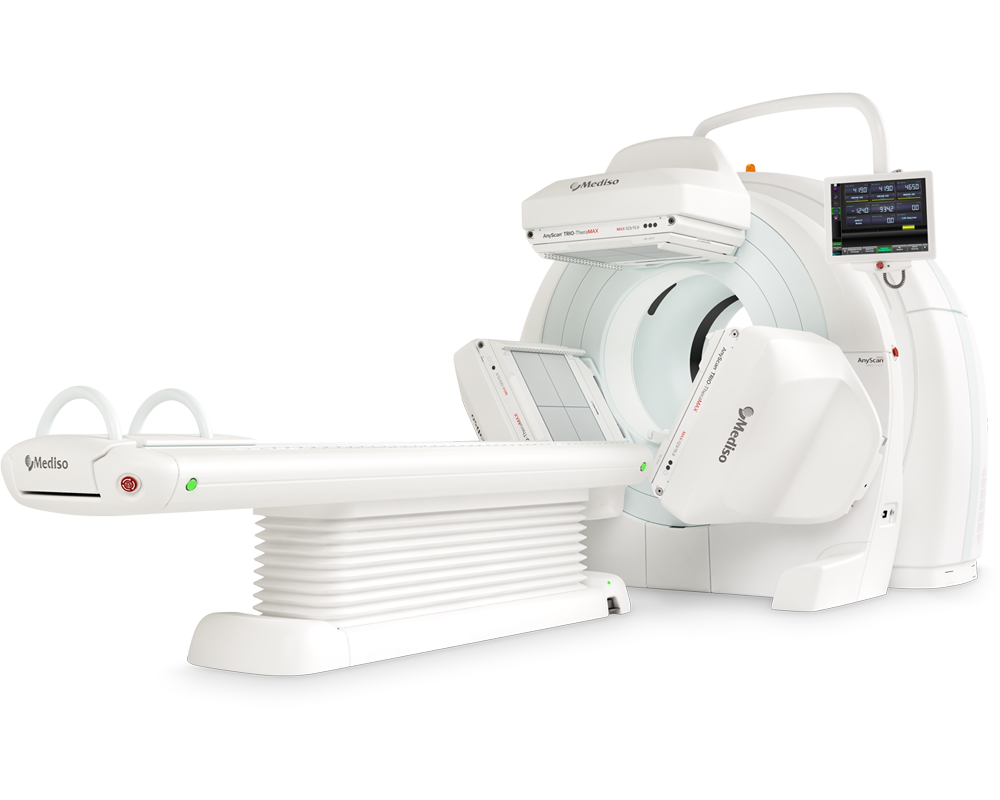
AnyScan® TRIO-TheraMAX SPECT/CT
Jakość Obrazów Teranostycznych i Diagnostycznych na MAXymalnym poziomie


W czym możemy pomóc?
Skontaktuj się z nami aby uzyskać informacje techniczne i / lub wsparcie dotyczące naszych produktów i usług.
Napisz do nas