Diagnostic value of [99mTc]Tc-PSMA-I&S-SPECT/CT for the primary staging and restaging of prostate cancer
2024.01.18.
Farkas et al., Therapeutic Advances in Medical Oncology, 2024
Introduction
A large number of studies have proved that prostate-specific membrane antigen-positron emission tomography/computer tomography (PSMA-PET/CT) provides excellent accuracy in primary staging and restaging of prostate cancer. Less data exist with PSMA-single photon emission computed tomography (SPECT)/CT investigations. The aim of this study was to evaluate the performance of [99mTc]Tc-PSMA-I&S (for imaging and surgery) in prostate cancer.
Methods
Study population
We examined 21 healthy volunteers and 100 male patients with histologically confirmed prostate cancer.

Follow-up
After PSMA-SPECT/CT, patients were followed up for at least 6 months. Results of the histopathological report, focal radiation therapy response assessment by morphological [multiparametric magnetic resonance imaging (MRI), CT] and functional (bone scintigraphy) imaging methods and PSA monitoring were used. Patients were contacted regularly to obtain follow-up information.
PSMA-SPECT/CT image acquisition protocol and image analysis
Mean activity of 666 ± 102 MBq [99mTc]Tc-PSMA-I&S (Institute of Isotopes Co. Ltd, Budapest, Hungary) was administered intravenously. Prior to imaging, patients were given an oral contrast agent (1000 mL of polyethylene glycol solution) to drink continuously, starting 60 min before the examination to promote bowel distention without introducing contrast material-induced attenuation correction artefacts. No adverse effects were observed in any of the patients after tracer injection. All scans were performed on an integrated whole-body SPECT/CT system (Mediso AnyScan® TRIO SPECT/CT; Mediso Medical Imaging Systems Ltd, Budapest, Hungary). Whole body SPECT imaging was carried out 6 h after the administration of the radiopharmaceutical (360°; 96 projections, 10 s/frame, matrix: 128 × 128, pixel: 4.22 mm). SPECT data collection was completed by low-dose CT (120 kV and 70 mAs) acquisition. Head–neck region imaging is conducted with a single field of view (FOV) measuring 40 cm. For torso imaging, the range extends from the supraclavicular region down to the mid-thigh, encompassing 2–3 FOVs (2–3 × 40 cm), with an overlap of at least 5 cm. The choice of 2 or 3 FOVs depends on the height of the patient.
PSMA-SPECT/CT images were visually evaluated for the presence of pathological radiopharmaceutical uptake by consensus reading of two nuclear medicine physicians with at least 20 years of experience. Physiological PSMA ligand uptake was found in the lacrimal gland, salivary glands, liver, spleen, small intestine, colon and kidneys. Patients were considered positive in the presence of markedly increased prostatic radiotracer uptake or at least one focal area of non-physiological PSMA expression in any region on SPECT scans.
The PSMA expression of pathological lesions was assessed by measurement of the maximum activity concentration [kBq/mL] in attenuation-corrected reconstructed images in each patient. The radiopharmaceutical uptake of the whole prostate gland in healthy volunteers was also measured.
As a reference, we used the mean activity concentration of gluteal muscles, aortic arch and healthy liver tissue. To obtain these values, at least 30 cm3 of volume of interest (VOI) was defined on SPECT scans without any visible abnormalities on CT.
For patients with biochemical recurrence or progressive disease, detection rates were calculated, defined as the proportion of scans with at least one PSMA-positive lesion.
Results
Primary prostate cancer
Patient-based diagnostic performance for detecting primary prostate cancer is as follows: sensitivity, specificity, positive predictive value, negative predictive value and accuracy of PSMA-SPECT/CT was 86%, 100%, 100%, 83% and 92%, respectively.

MRI and PSMA-SPECT/CT scan of a 59-year-old patient with primary prostate cancer (ISUP V/V.; PSA 19.20 ng/mL). The left posterolateral peripheral zone of the prostate demonstrates 16 mm mass with increased signal intensities (a) on the DWI images with focal ADC decrease (b) and decreased signal intensities on the T2w images (c), PSMA-SPECT/CT images (d) show increased radiopharmaceutical uptake in the same region (red arrowheads).
Primary staging (N, M)
In patient group 1, patient-based diagnostic performance is as follows: sensitivity, specificity, positive predictive value, negative predictive value and accuracy of PSMA-SPECT/CT for detecting metastases in primary staging were 88%, 100%, 100%, 85% and 93%, respectively.
Biochemical recurrence and restaging
In patient group 2, PSMA-SPECT/CT patient-based sensitivity was 72%.
In all, 12 patients received salvage radiotherapy to the prostate bed with pelvic lymph node regions and 5 patients received systemic treatment after a PSMA scan. In these patients, local recurrence or pelvic lymph node involvement could not be clearly distinguished only metastatic involvement could be confirmed (one case) so data from these patients were not included in the following calculations. This way, patient-based sensitivity, specificity, positive predictive value, negative predictive value and accuracy for local recurrence were 67%, 100%, 100%, 86% and 89%, and for detecting metastases 91%, 92%, 98%, 75% and 91%, respectively.

PSMA SPECT/CT – axial (a–c); coronal (d) and sagittal slices (e) – scan of 82-year-old metastatic prostate cancer patient (ISUP V/V.; PSA: 618 ng/mL) with newly diagnosed liver lesions by ultrasound. The PSMA SPECT/CT proved high PSMA expression in the hepatic lesions indicative of prostate cancer metastases. Multifocal bone metastases on the spine and pelvic bone and several moderately enlarged lymph nodes with high PSMA density were shown as well. Total liver tumour burden 320 mL.
Discussion
In this retrospective single-centre study, [99mTc]Tc-PSMA-I&S was used for detecting primary prostate cancer, primary staging and restaging in biochemical recurrence or case of suspected disease progression. Although PSMA-I&S was originally developed for radio-guided surgery purposes, it was found to be a suitable radiotracer for prostate cancer imaging, as 6 h post-injection scans showed high contrast and proper image quality. There are some advantages of PET/CT compared to SPECT/CT: PET/CT typically offers slightly higher spatial resolution (dependent on the specific devices used) and shorter uptake times (in this case 6 versus 1–2 h). During the current examination, the acquisition time averaged 35 min (median), which is comparable to the duration of most PET examinations commonly employed in general practice, though it may be longer than what is achieved with more recent PET camera technology.19
This technique can be utilized in nuclear medicine departments where PET/CT or PSMA-PET tracers are not available. Simple and reliable radiolabelling procedures facilitate the use of this radiopharmaceutical so PSMA-SPECT/CT can be easily integrated into routine diagnostic practice.
Full article on Therapeutic Advances in Medical Oncology
-
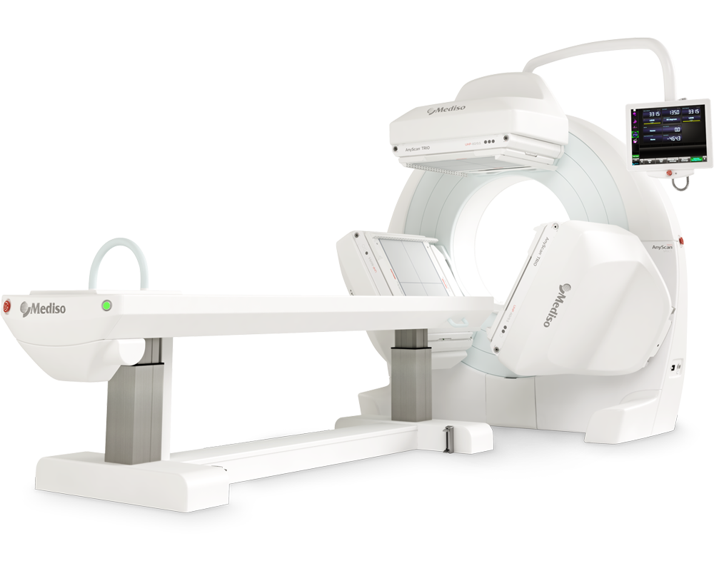
AnyScan® TRIO SPECT
Ultra-fast Triple-NaI-Detector SPECT for wide range of clinical applications
-
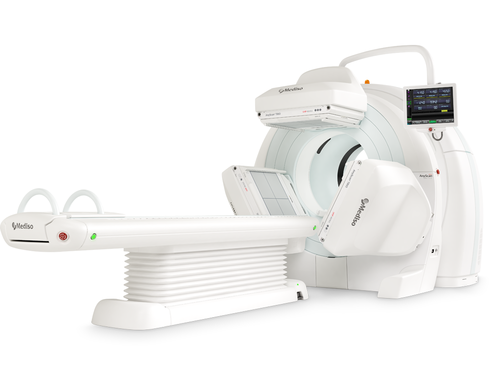
AnyScan® TRIO SPECT/CT
Ultra-fast Triple-NaI-Detector SPECT system for wide range of clinical applicati...
-
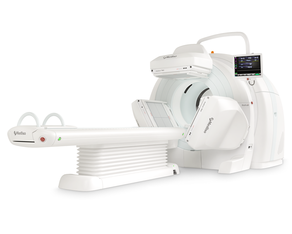
AnyScan® TRIO SPECT/CT/PET
An integrated triple-detector SPECT/CT and PET/CT system in a single-room instal...
-
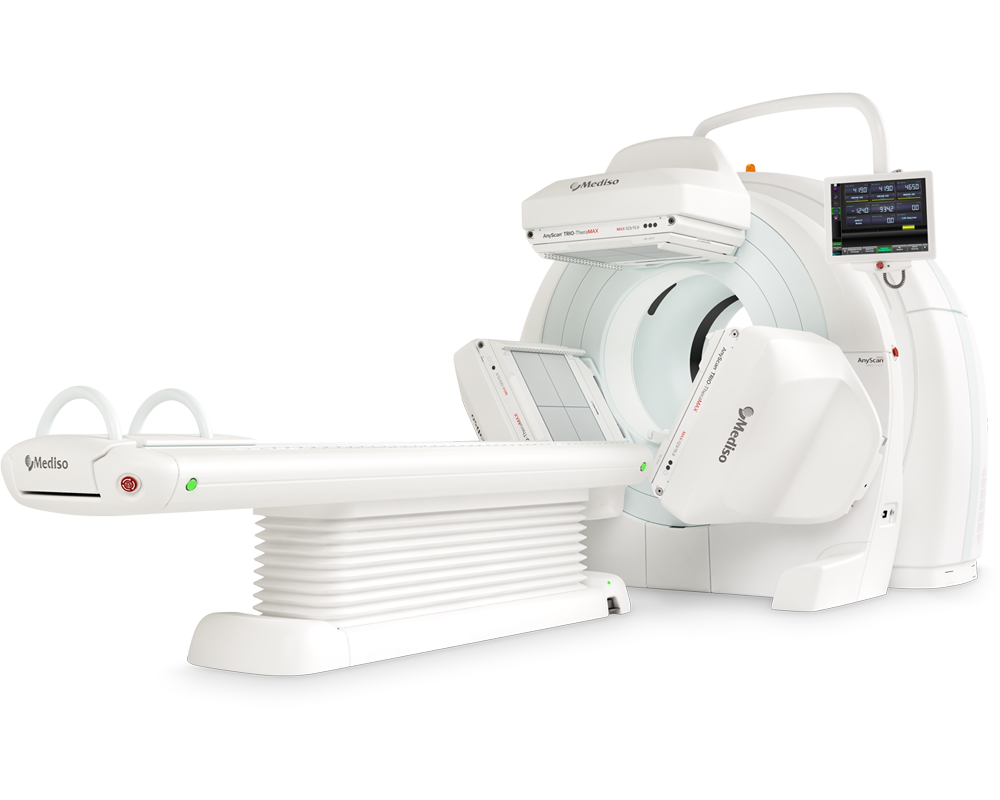
AnyScan® TRIO-TheraMAX SPECT/CT
Jakość Obrazów Teranostycznych i Diagnostycznych na MAXymalnym poziomie


W czym możemy pomóc?
Skontaktuj się z nami aby uzyskać informacje techniczne i / lub wsparcie dotyczące naszych produktów i usług.
Napisz do nas