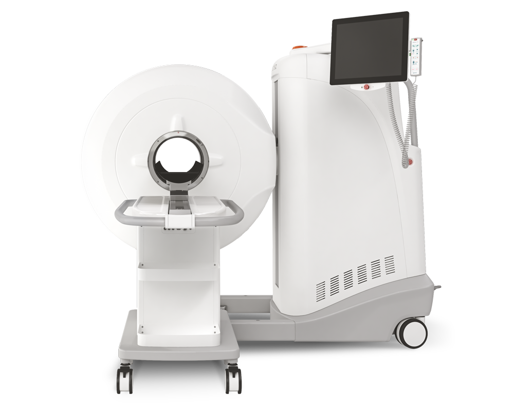
Refined acquisition of high-resolution chest CTs in macaques by free breathing
2021.07.02.
Botond Tölgyesi et al., Laboratory Animals, 2021
Abstract
The use of medical imaging as a non-invasive or minimally invasive method to assess disease or treatment response continues to grow. A similar trend is observed in pre-clinical research, in general, and more specifically in macaques, enabling longitudinal assessment of disease in individual animals. Computed tomography (CT) is such an imaging technique used to obtain clinically applicable data. To acquire a chest CT using a cone beam tomography system, some kind of respiration control is needed. A commonly used technique for this is endotracheal intubation and mechanical ventilation. However, although routinely performed this can increase the risk of impact on welfare in comparison with non-invasive imaging. Therefore, we studied the option of retrospectively gated CTs: acquiring high resolution chest CTs in freely breathing macaques. For this, we compared 748 CTs obtained during free breathing with 881 CTs obtained with mechanical ventilation in combination with a breath-hold procedure predominantly on the appearance of misregistration artifacts. The scans were obtained during different stages of multiple experimentally induced respiratory diseases. The comparison shows that although there are still streaking artifacts present in the retrospective gated scans, the amount of shading artifacts is reduced to reveal the proper diagnosis in SARS-CoV-2 in example. The authors' data reveal that the use of retrospective gating in high resolution CTs for macaques can be successfully applied. With the use of this technique, artifacts due to free breathing are reduced to a diagnostically appropriate level. Most importantly, this technique makes chest CTs with this instrumentation a non-invasive modality.
Results from MultiScan™ LFER PET/CT
The image quality of the optimized retrospective gating reconstruction was visually compared with a free breathing and a breath-hold procedure in Fig 5a-c. Images were obtained from the same animal within 2 days of each other. A free breathing image was reconstructed as part of the gated scan. Besides the comparison of the three imaging techniques with healthy macaques, two examples of infected animals are also shown (Fig. 6). Fig. 6a,c shows an example of a rhesus macaque 6 weeks after the first infection with Mycobacterium tuberculosis following a repeated low dose scheme of 8 weeks 1 colony forming unit (CFU) with two consolidations in the lower left part of the lung. Fig. 6b,d shows an example of a rhesus macaque 7 days after severe acute respiratory syndrome coronavirus 2 (SARS-CoV-2) infection with a ground glass opacity (GGO) in the lower dorsal part of the right lung.

Fig. 5.: Free breathing with retrospective gating (a), free breathing without retrospective gating (b) and breath-hold (c) coronal and transversal representative CT slices of one rhesus macaque (Macaca mulatta).

Fig. 6.: Free breathing with retrospective gating (a and b) and free breathing without retrospective gating (c and d) sagittal CT slices of two rhesus macaques (Macaca mulatta) after infection with mycobacterium tuberculosis (a and c) or SARS-CoV-2 (b and d). The cross hairs in the image mark the lesion.
Discussion
This report demonstrates the effectiveness of retrospective respiratory gating in improving high resolution CT images from free breathing macaques. To evaluate the effectiveness of the gating procedure, one has to compare it with the other two options: free breathing, and some type of respiratory control, in this case a breath hold procedure.
Provided that the captured data is of sufficiently high quality, imaging can address the 3 rotations by providing refinement as longitudinal information is obtained in a minimal or non-invasive way that reduces procedure severity. Nevertheless, this refinement can be shattered by reduced image quality. Image quality of a CT is, for the most part, determined by the number of artifacts present. Respiratory motion artifacts, so called misregistration artifacts, cause blur, shading and streaking on the reconstructed image. Streaking appear as straight lines, bright and dark, across the image. Closer to the bones (vertebrae, ribs) the streaks intensify. In general, these artifacts are unlikely to cause misdiagnosis, since pathology rarely appears like this but they reduce the reliability of the results. Streaks are present in both the free breathing and gated reconstruction though their intensity is reduced with gating. Shading artifacts show often in regions of high contrast (e.g. between soft-tissue and bones or soft tissue and air), causing shifts in HU representations. This artifact is less predictable compared with streaks, and the boundaries are less easy to identify since natural anatomy also presents gradual changes in HUs. As a result, shading can mimic pathology on it is own or dominate underlying lesions. This last point can lead to misdiagnosis; for instance, in COVID-19 research, the most common lesion types found after SARS-CoV-2 infection on CT are GGOs, which are characterized by only a slight increase in lung opacity. The risk of missing these types of lesions is visualised in Figure 6, disturbing the image quality in a clinical relevant way.
- When comparing the three breathing techniques, shading is totally absent in the breath-hold image and almost absent in the gated image compared with the free breathing image when focussing on the diaphragm and trachea. The findings of this study are likely to be translated to other animal species, including other non-human primate species, when respiratory control is necessary. In humans, for certain CT protocols a similar type of retrospective gating is applied.
- The data of the authors show that the use of retrospective respiratory gating in high resolution CTs for macaques is successful. With the use of this technique, artifacts due to free breathing are reduced to a diagnostically appropriate level. Most importantly, this technique makes chest CTs with this instrumentation a non-invasive modality.
W czym możemy pomóc?
Skontaktuj się z nami aby uzyskać informacje techniczne i / lub wsparcie dotyczące naszych produktów i usług.
Napisz do nas