Gadolinium-Based Nanoparticles Sensitize Ovarian Peritoneal Carcinomatosis to Targeted Radionuclide Therapy
2023.10.19.
Clara Diaz Garcia-Prada et al., Journal of Nuclear Medicine, 2023
Summary
Ovarian cancer (OC) stands as the deadliest among gynecologic malignancies, with a 5-year overall survival rate of 46%. Typically, OC is identified at a stage where it has already disseminated to the peritoneal cavity, leading to peritoneal carcinomatosis. This research delved into the potential augmentation of targeted radionuclide therapy efficacy through the utilization of gadolinium-based nanoparticles (Gd-NPs) in conjunction with [177Lu]Lu-DOTA-trastuzumab an antibody designed to combat the human epidermal growth factor receptor 2. Gd-NPs exhibit radiosensitizing effects, a trait observed in traditional external-beam radiotherapy and validated through clinical phase II trials.
Results from the SPECT-CT
Initially, a calibration curve was established that correlated the bioluminescence signal with the tumor mass. This curve facilitated tumor growth monitoring, optimal treatment initiation time, and end point determination in euthanized mice with SK-OV-3-luc cell xenografts in the peritoneal carcinomatosis (OC-PC) model of ovarian cancer. Specifically, euthanasia was performed when the bioluminescence signal reached 4 × 10^10 photons/s, corresponding to a total tumor mass of approximately 2000 mg.
Gd-NP and [177Lu]Lu-DOTA-Trastuzumab Biodistribution in Mice
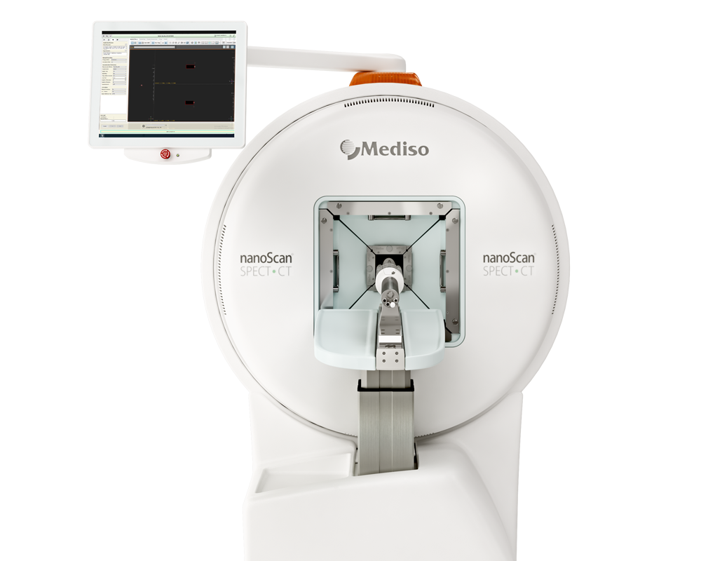
In the biodistribution study involving mice carrying SK-OV-3-luc cell xenografts, the uptake pattern of [111In]In-DTPA-trastuzumab in tumors reached its zenith between 24 and 72 h post-injection, peaking significantly at 48 h (71.5% ± 7.7% injected activity/g) (Fig. 1, left). Meanwhile, blood uptake showed its peak at 24 h (17.9% ± 2.4% injected activity/g), gradually diminishing to 17.9% ± 2.4% at 168 h post-injection. Conversely, Gd-NPs exhibited maximal tumor uptake at 30 min post-injection (4,267 ± 80 mmol/g), decreasing to 804 ± 321 mmol/g at 6 h (Fig. 1, middle). Notably, Gd-NPs accumulated prominently in the kidneys (5,138 ± 317 mmol/g) at 6 h, indicating renal elimination. SPECT/CT (Mediso Ltd.) imaging following intraperitoneal injection of [111In]In-Gd-NPs affirmed tumor uptake and renal excretion (Supplemental Fig. 1B). Furthermore, ex vivo dual-isotope SPECT/CT (Mediso Ltd.) imaging validated the colocalization of [177Lu]Lu-Gd-NPs and [125I]I-trastuzumab in collected tumor nodules from mice (Fig. 1, right). Consequently, in evaluating the combination therapy, animals received a single injection of [177Lu]Lu-DOTA-trastuzumab, followed by Gd-NPs after a 48-hour interval.

FIGURE 1. (Left) Biodistribution of [111In]In-DTPA-trastuzumab determined by ex vivo γ-counting of tumor nodules and organs collected at various times (4 per time point) after intraperitoneal injection. (Middle) Biodistribution of Gd-NPs by inductively coupled plasma mass spectrometry in tumor nodules and organs collected at various times after intraperitoneal injection (3 per time point). Results are mean ± SEM. (Right) Ex vivo SPECT/CT dual-isotope imaging of SK-OV-3-luc cell tumors collected from mice after intraperitoneal injection of [125I]I-trastuzumab and [177Lu]Lu-CuPRiX (NH TherAguix S.A. and Institut Lumière Matière) Gd-NPs. Merged images confirmed colocalization of trastuzumab and CuPRiX NPs. % IA = percentage injected activity; DTPA = diethylenetriaminepentaacetic acid; ICP-MS = inductively coupled plasma mass spectrometry; trastu = trastuzumab.
In the initial series of experiments evaluating the combination of [177Lu]Lu-DOTA-trastuzumab and Gd-NPs, mice were euthanized at 4 weeks post-treatment. Tumor weight was significantly reduced in mice treated with 50 µg trastuzumab and 10 µg Gd-NP compared to the NaCl-treated group (P = 0.0150). In addition, treatment with [177Lu]Lu-DOTA-trastuzumab at maximal tolerable activity (10 MBq) significantly reduced tumor weight at week 4, and the addition of 10 mg Gd-NPs resulted in a further significant reduction compared to trastuzumab plus Gd. NPs (P = 0.0030). However, the efficacy of [177Lu]Lu-DOTA-trastuzumab was not significantly increased by the addition of Gd-NPs (P = 0.3788). Consequently, subsequent experiments with reduced [177Lu]Lu-DOTA-trastuzumab activity (5 MBq and 2.5 MBq) showed that 5 MBq maintained therapeutic activity, whereas 2.5 MBq was insufficient to observe further therapeutic effects of 177Lu irradiation, with a higher tumor mass than in comparison for the 5 MBq group (P = 0.0040).
Hence, the optimal injected activity was determined to be 5 MBq, striking a favorable balance between efficacy and the potential radiosensitizing effect mediated by Gd-NPs
In the study of the fractionation regimen, which aimed to enhance the therapeutic efficacy of targeted radionuclide therapy (TRT), mice were anesthetized 4 weeks after xenograft. The combination of [177Lu]Lu-DOTA-trastuzumab (5 MBq) and Gd-NPs, given according to the 3rd dosing regimen (2 × 5 mg/day at 24 and 72 h after TRT) showed the most significant reduction in tumor mass (P) showed. = 0.0320) compared to treatment with [177Lu]Lu-DOTA-trastuzumab alone. In addition, regimen 3 showed a significant improvement in objective response rate and median survival, superior to other treatment groups. These results suggest that fractional administration of Gd-NPs, especially in scheme 3, enhances the efficacy of TRT in ovarian cancer.

FIGURE 3. Kaplan–Meier survival analysis of mice bearing intraperitoneal SK-OV-3-luc tumor cell xenografts. Mice received single intraperitoneal injection of NaCl, 25 μg of trastuzumab plus Gd-NPs (2 × 5 mg per day, 24 and 72 h after trastuzumab), 5 MBq of [177Lu]Lu-DOTA-trastuzumab, or 5 MBq of [177Lu]Lu-DOTA-trastuzumab plus Gd-NPs (2 × 5 mg per day, 24 and 72 h after TRT). R3 = regimen 3.
Upon administering 5 MBq of [177Lu]Lu-DOTA-trastuzumab, the mean absorbed dose in tumors reached 1.99 ± 0.21 Gy, while normal tissues exhibited doses less than 0.5 Gy, with the exception of blood (0.570 ± 0.002 Gy) (Supplemental Fig. 5). These findings were crucial for evaluating the potential contribution of Gd-NPs to absorbed dose enhancement.
To explore the in vivo radiosensitizing effect of Gd-NPs, the cellular uptake of Gd-NPs (10 mg/mL) in SK-OV-3-luc cells was monitored at various time points (2 to 144 h). The uptake plateaued between 6 and 18 h (0.047 ± 0.003 pg/cell) before slowly decreasing. Gd-NPs, detected in the cytoplasm but not in the cell nucleus, colocalized with lysosomes but not mitochondria. [177Lu]Lu-DOTA-trastuzumab uptake peaked at 18 h (0.045 ± 0.007 Bq/cell) before gradually decreasing. Clonogenic survival analysis of SK-OV-3-luc, A-431, and OVCAR-3 cells exposed to [177Lu]Lu-DOTA-trastuzumab, with or without Gd-NPs (10 mg/mL), revealed a radiosensitizing effect, achieving the same cytotoxic effect with half the tested activity. Bliss independence model and oxidative stress inhibition experiments confirmed the synergistic and oxidative stress-dependent radiosensitizing effect of Gd-NPs.

FIGURE 6. Oxidative stress is involved in radiosensitizing effects of Gd-NPs. (A) SK-OV-3-luc cells were incubated with [177Lu]Lu-DOTA-trastuzumab, 1 MBq/mL, for 18 h with or without Gd-NPs, 10 mg/mL, with or without N-acetyl-l-cysteine (NAC), catalase, or dimethyl sulfoxide. Then, clonogenic survival was measured. (B) Clonogenic survival of SK-OV-3-luc, A-431, and OVCAR-3 cells incubated with [177Lu]Lu-DOTA-trastuzumab (0–4 MBq/mL) with or without Gd-NPs, 10 mg/mL, for 18 h in presence or not of DFP. Results are mean ± SD of 3 independent experiments performed in triplicate. *P < 0.05. **P < 0.01. ***P < 0.001. ****P < 0.0001. DFP = deferiprone; DMSO = dimethyl sulfoxide; NAC = N-acetyl-l-cysteine; ns = not significant (Mann–Whitney t test) compared with cells treated with [177Lu]Lu-DOTA-trastuzumab.
Generation of Auger Electrons Through Gd-NP Exposure to [177Lu]Lu-DOTA-Trastuzumab
Monte Carlo simulations were employed to explore the interaction between 177Lu atoms and lysosomes, considering scenarios with and without Gd-NPs. The simulations involved two steps (Supplemental Fig. 8): in the first step, 177Lu atoms were distributed within a cell or in the 1-mm-diameter medium surrounding the cell, undergoing radioactive decay. In the second step, particles from the phase space were simulated in lysosomes with a variable number of Gd-NPs, based on uptake assay results. The continuous energy spectrum (0–498 keV) of electrons emitted by 177Lu in lysosomes showed a peak around 100 keV. Dose rates and accumulated doses in lysosomes were evaluated at different time points for incubation and postincubation scenarios. Results indicated that Gd-NPs did not contribute to increased absorbed dose, and the contribution of photoelectrons was minimal. However, the presence of Gd-NPs resulted in an elevated production rate and cumulative number of Auger electrons in lysosomes under the same conditions and scenarios (Fig. 7).

FIGURE 7. Physical aspects of TRT combined with Gd-NPs. Dose rate and accumulated dose in lysosomes at various time points for incubation and postincubation scenarios (left). Auger electron production rate and cumulative number in lysosomes under same conditions as described for left panel (right).
Conclusion
This pioneering study is the first to demonstrate the synergistic impact of combining Targeted Radionuclide Therapy (TRT) with Gadolinium-based nanoparticles (Gd-NPs), achieving radiosensitization and enhanced therapeutic efficacy with reduced injected activity. These findings offer a promising avenue for minimizing radiation-induced toxicities in patients. The proposed combination is particularly attractive for adjuvant TRT post-cytoreductive surgery, and its clinical translation is facilitated by the existing use of Gd-NPs in conjunction with External Beam Radiation Therapy (EBRT). Exploring intravenous administration is a pertinent consideration, addressing potential complications associated with intraperitoneal delivery methods, such as infection risks linked to indwelling catheters in clinical trials for peritoneal metastases.
Full article on jnm.snmjournals.org
W czym możemy pomóc?
Skontaktuj się z nami aby uzyskać informacje techniczne i / lub wsparcie dotyczące naszych produktów i usług.
Napisz do nas