Development of [18F]LU14 for PET Imaging of Cannabinoid Receptor Type 2 in the Brain
2021.07.28.
Rodrigo Teodoro et al., International Journal of Molecular Sciences, 2021
Summary
The psychoactive effect of the well-known drug marijuana is caused by the active ingredient (−)-Δ9-trans-tetrahydrocannabinol (THC), which abundantly occurs in the cannabis plant. The investigation of the mechanism through which THC manifests its biological activity led to the discovery of the endocannabinoid system. It comprises a class of transmembrane proteins that belongs to the superfamily of G-protein-coupled receptors; their endogenous ligands (endocannabinoids), the endocannabinoid-synthesising and -degrading enzymes, as well as the endocannabinoid transporters. Two types of cannabinoid receptors, namely cannabinoid receptors type 1 (CB1R) and cannabinoid receptors type 2 (CB2R), were identified so far and are being thoroughly investigated. Both CB1R and CB2R transduce signals from extracellular to intracellular space by inhibiting the activity of adenylyl cyclase over Gi/o proteins, thereby suppressing the subsequent cyclic adenosine monophosphate (cAMP) pathway. While the CB1R is associated with the central nervous system and is abundantly expressed in the brain (highest density in the hippocampus, cerebellum and striatum), the CB2R is found in the spleen, tonsils and the thymus gland and modulates immune cell functions. In the brain, the CB2R are present at low levels, and their overexpression is associated with neurodegenerative diseases such as Alzheimer’s disease, Parkinson’s disease, Huntington’s disease and cancer.
More recently, it has been shown that CB2R is expressed in dopaminergic neurons in the midbrain and, in glutamatergic neurons in the red nucleus, modulates motor behaviour in mice. Others have shown that the selective activation of the CB2R receptor results in cell apoptosis, the inhibition of tumor cell growth and the inhibition of neo-angiogenesis. Moreover, selective CB2R agonists were successfully used to suppress inflammatory and neuropathic pain without psychotropic side effects caused by the use of THC or other CB1R-related pharmaceuticals. The overexpression of CB2R receptors was correlated with microglial polarisation from a pro-inflammatory M1 to anti-inflammatory M2 state by a yet not fully elucidated mechanism. Thus, CB2R are regarded as a highly valuable marker for the early detection and treatment of several neuropathological conditions. The investigation of CB2R as a target to develop novel therapeutic and early diagnostic strategies gained importance over the past years. The non-invasive imaging of CB2R with positron emission tomography (PET) is of great interest, as demonstrated by the large number of positron-emitting radioligands developed over the past years.
Despite tremendous efforts, the development of a suitable radiotracer for PET imaging of the CB2R in the brain remains a challenging task. To gain eligibility for consideration as a potential PET radioligand for brain imaging, the first criterion to be fulfilled by a CB2R ligand is high receptor affinity and selectivity, which in many cases is hampered by the homologous similarity between CB1R and CB2R. Another important criterion is a balanced lipophilicity which is usually used as a predicting tool for (1) the ability to cross the blood–brain barrier and (2) non-specific binding.
In this work, the authors focus on the development and biological evaluation of a radiofluorinated compound belonging to the naphthyridin-2-one class, and they selected compound 5 due to the published high affinity and selectivity towards CB2R (Ki (hCB2R) = 1.3 nM, SI > 700) and its favourable physicochemical properties for targeting the brain. In order to evaluate in vivo the ability of LU14 to image CB2R in the brain by PET, the authors selected a well-established rat model carrying an adeno-associated viral (AAV2/7) vector expressing hCB2R(D80N) at high densities in a striatal region.
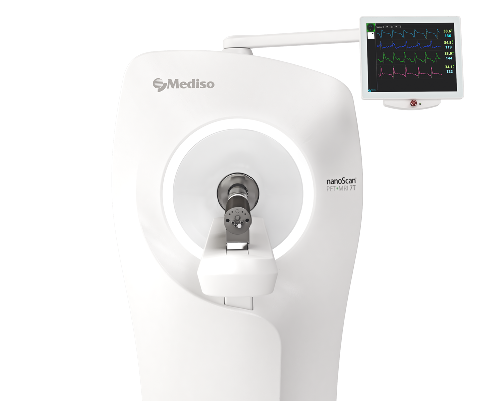
Results from nanoScan PET/MRI 1T
In vivo biodistribution of [18F]LU14 in rats was assessed by dynamic small animal PET 60 min recordings, followed by T1-weighted (GRE, TR/TE = 15.0/2.4 ms, 252/252, FA = 25°) MR imaging with whole-body coils for anatomical correlation and attenuation correction. For the uptake studies into the spleen three male Wistar Hun rats weighing 268 ± 56 g were used. For the uptake studies into the brain, five female Wistar rats weighing 263 ± 13 g carrying the stereotactically injected AAV 2/7-CaMKII0.4-intron-hCB2R D80N (right striatum) and AAV2/7-CaMKII0.4-intron-3flag-eGFP (contralateral, left striatum). Animals were initially anaesthetised with 5% isoflurane and placed on a thermostatically heated animal bed where anaesthesia was maintained with 2% isoflurane in 60% oxygen/38% room air. They were pre-treated by i.v. injections of vehicle solution only, containing DMSO: Kolliphor EL: saline in a composition of 1:2:7 (control group) or the CB2R agonist GW405833 (1.5 mg/kg bodyweight for spleen-scan experiments and 5 mg/kg for the displacement experiments with local overexpression of hCB2R) 10 min prior [18F]LU14 administration. [18F]LU14 (22.4 ± 3.5 MBq) was injected i.v. into the lateral tail vein (bolus within 5 s) at the start of the PET acquisition. List-mode PET data were binned as a series of attenuation-corrected sinogram frames (12 × 10 s, 6 × 30 s, 5 × 60 s and 10 × 300 s) and were reconstructed by ordered subset expectation maximisation (OSEM3D) with four iterations, six subsets and a voxel size of 0.4 mm3. The analysis of reconstructed datasets was performed with PMOD software (v4.103, PMOD Technologies LLC, Zurich, Switzerland).
In order to evaluate the ability of [18F]LU14 to penetrate the blood–brain barrier and potentially image the human CB2R (hCB2R) in pathological conditions, PET imaging experiments were performed with [18F]LU14 in a rat model with local overexpression of hCB2 receptors in the right striatum, as described by Vandeputte et al. and validated as a suitable model for evaluation of both CB2R agonists and antagonists by Attili. As shown in Figure 6, analysis of the TACs revealed a significantly higher uptake into the striatal region overexpressing the hCB2R(D80N), compared to the contralateral region and the cerebellum after i.v. injection of [18F]LU14. The initial uptake was followed by a washout with a significantly longer mean residence time (MRT) in the target region compared to non-target regions.
The SUVr (hCB2R-to- contralateral ratio) reached a plateau (6.6 ± 1.7 and 7.0 ± 1.7) at 45 min p.i. and the SUVr (hCB2R-to-cerebellum ratio) an earlier plateau (5.8 ± 1.1 to 6.5 ± 1.4) at 30 min p.i. Thus, both reference regions demonstrated favourable kinetics of this tracer to image cerebral CB2R. The uptake of [18F]LU14 in the brain region with CB2R overexpression was about five-fold higher compared to the previously reported radioligand [18F]MA3 and about two-fold higher compared to [11C]NE40 in the same rat model. In order to assess whether the binding of [18F]LU14 to the CB2R is specific and reversible, displacement experiments were performed by the intravenous administration of the known CB2R agonist GW405833 at 20 min p.i (Figure 6B). The amount of activity in the brain region with CB2R overexpression was considerably displaceable by the competitor, as shown by the significant reduction of the AUC20–60min by 73% (compared to the contralateral region) and 76% (compared to the cerebellum).
In summary, these experiments demonstrate that [18F]LU14 is a specific CB2R radioligand with reversible receptor binding, favourable kinetics and high potential to be used for the detection of cerebral CB2R overexpression in pathological conditions like neuroinflammation or brain cancer.

- These experiments confirm the ability of [18F]LU14 to penetrate the BBB
- The model clearly reveals a specific uptake of [18F]LU14 under pseudopathological conditions of elevated hCB2R expression
- Due to the negligible uptake of radioactivity in the cerebellum after i.v. of [18F]LU14 during the time span of the analysis, this region could be used as a reliable reference region in this analysis.
W czym możemy pomóc?
Skontaktuj się z nami aby uzyskać informacje techniczne i / lub wsparcie dotyczące naszych produktów i usług.
Napisz do nas
