Design, Radiosynthesis and Preliminary Biological Evaluation in Mice of a Brain-Penetrant 18F-Labelled σ2 Receptor Ligand
2021.05.21.
Rareş-Petru Moldovan et al., International Journal of Molecular Sciences, 2021
Summary
The presence of two distinct classes of σ receptors, which was postulated already in the 1970s based on the effects of opiate benzomorphans, was recognized two decades later. Differences in the stereoselectivity, pharmacology, and molecular weight of guinea pig brain σ receptors and the binding sites of σ receptor ligands in PC12 cells, a tumor cell line derived from rat adrenal chromaffin cells, led to the designation of σ1 and σ2 receptors, respectively. By contrast to the structurally and functionally thoroughly investigated and well described σ1 receptor (for recent reviews of structure, function, and therapeutic potential, a profound characterization of the biology and a comprehensive understanding of the therapeutic potential of the σ2 receptor were delayed mainly due to the absent identification of the protein and the coding gene. A significant step forward was the identification of progesterone receptor membrane component 1 (PGRMC1) as part of the protein complex containing the binding site of σ2 receptor ligands, which was developed in 2011. Further detailed analyses of the relation between PGRMC1 and the σ2 receptor eventually resulted in the identification of the σ2 receptor as the transmembrane protein 97 (TMEM97), singled out by radioligand binding characteristics from candidate proteins initially isolated via σ2 receptor ligand affinity chromatography from calf liver.
The elucidation of the molecular identity of the σ2 receptor and the identification of this protein as part of a functional structure regulating the cholesterol homeostasis of cells significantly improved the understanding of the role of the σ2 receptor and the therapeutic potential of σ2 receptor ligands for neurologic and neurodegenerative disorders as well as cancer.
A much higher expression of σ2 receptors in cancer cells in comparison to normal cells revealed by radioligand binding studies in the 1990s led immediately to the evaluation of the potential of σ receptor-targeting radiopharmaceuticals for noninvasive imaging of human malignancies and to the postulation of the potential of σ2 receptors as biomarkers of the proliferative status of solid tumors. Besides, particular ligands targeting the σ2 receptor have the potential to activate the apoptotic program in cancer cells. The current classification of the σ2 receptor ligands as agonists or antagonists is mainly based on this apoptotic potential, with pro-apoptotic ligands such as siramesine designated as agonists.
While to the best of our knowledge none of the selective agonists of the σ2 receptor has been tested in clinical treatment studies, a number of structurally different radiolabeled σ2 receptor ligands have been developed and applied as molecular imaging probes in preclinical and clinical positron emission tomography (PET) studies, such as [11C]1, [18F]2, [76Br]3, [76Br]4, [11C]5–8, [18F]9 or [18F]10, called [18F]ISO-I and its derivatives [18F]11, [18F]12, and [18F]13. As deduced earlier from a substantial and local uptake of [11C]RHM-1 in glioblastoma in an orthotopic mouse model, the correlation between the uptake of [18F]ISO-I in the tumors of patients with lymphoma, breast cancer, and head and neck cancer and the expression of the proliferation marker Ki-67 supported the suitability of quantitative noninvasive imaging of σ2 receptors by PET for monitoring of the proliferative potential of cancer. In addition, a currently recruiting study (NCT03057743) applying PET with [18F]ISO-I as baseline scan in patients with metastatic breast cancer is expected to yield data regarding the value of σ2 receptor expression as a prognostic biomarker.
The clinically extremely relevant issue of an improvement of the management of patients with brain cancer, however, may not profit from noninvasive imaging with [18F]ISO-I due to the limited blood–brain barrier permeability of this particular radiotracer. Thus, the authors intended to develop a brain-penetrant radiotracer based on the well described 18F-fluoroethoxy-substituted 2-(4-(1H-indol-1-yl)butyl)-6,7-dimethoxy-1,2,3,4-tetrahydroisoquinoline class of compounds (e.g., compounds [18F]14 and [18F]15) recently shown to have a significantly higher brain uptake in comparison to [18F]ISO-I (3–4% ID/g at 2 min p.i. vs. 0.76% ID/g at 5 min p.i.). Using those compounds as a starting point, the authors intended to further improve the applicability of the corresponding radiotracers for quantitative monitoring of σ2 receptors in the brain. To further optimize pharmacological parameters such as selectivity, metabolic stability, and lipophilicity, the authors synthesized a novel series of nine fluorinated derivatives that all bore the F-atom at the original indole or, as a novel scaffold in the medicinal chemistry of σ2 receptor ligands, azaindole (pyrrolopyridine). The azaindole scaffold was introduced to assess the effects of this bioisosteric ring system. To explore the potential of azaindole-substituted σ2 receptor ligands for in vivo brain imaging, the authors radiofluorinated RM273, the most suitable ligand out of the series developed herein, and examined the pharmacokinetics of [18F]RM273 through dynamic PET studies in mice as well as its binding parameters through brain cryosections of an orthotopic rat glioblastoma model.
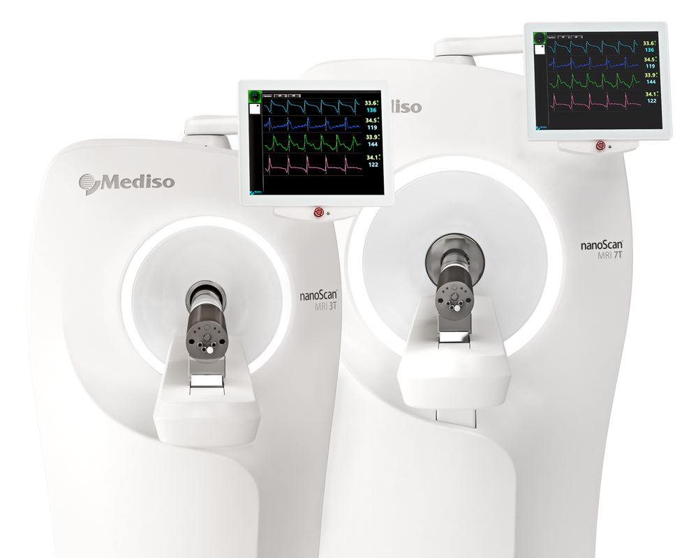
Results from nanoScan PET/MRI 1T
For the imaging studies, The biodistribution of [18F]RM273 in mice was assessed by dynamic small animal PET 60 min recordings, followed by T1 weighted (GRE, TR/TE = 15.0/2.4 ms, 252/252, FA = 25°) imaging for anatomical correlation and attenuation correction. The mice weighing 31.7 ± 3.8 g were anaesthetized with 2% isoflurane in 60% oxygen and placed on a thermostatically heated animal bed. [18F]RM273 (7.2 ± 1.1 MBq) was injected i.v. into the lateral tail vein (bolus within 5 s) at the start of the PET-acquisition. List mode PET data were binned as a series of attenuation-corrected sinogram frames (12 × 10 s, 6 × 30 s, 5 × 60 s and 10 × 300 s) and were reconstructed by Ordered Subset Expectation Maximization (OSEM3D) with four iterations, six subsets, and a voxel size of 0.4 mm3. The analysis of reconstructed datasets was performed with PMOD software. GraphPad Prism was used to calculate the area under the curve (AUC), as well as to determine the peak TAC half-life time by fitting of time–activity curves with dissociation one phase exponential decay setting t0 at peak of TAC.
As expected by the optimal lipophilicity of [18F]RM273 the PET images and the time–activity curve obtained for the whole brain shown in Figure 6. indicate a fast permeation of the BBB with a mean standardized uptake value (SUV) of 1.3 ± 0.4 at 2.25 min p.i. (n = 4). Furthermore, rapid clearance from the brain, with t1/2 of 13.1 min after peak time and a peak-to-endpoint ratio of 6.4 ± 0.9, suggests low non-specific binding of [18F]RM273 in vivo.
- The authors herein report the development of the PET radioligand [18F]RM273, which is suitable to monitor the expression of σ2 receptors in the brain.
- This new radioligand has the potential to support research on the validation of σ2 receptor expression as a biomarker of tumor-specific processes such as proliferation in cancers of the central nervous system.
- In addition, the development of therapies targeting σ2 receptors in the brain could be promoted by [18F]RM273 PET.
W czym możemy pomóc?
Skontaktuj się z nami aby uzyskać informacje techniczne i / lub wsparcie dotyczące naszych produktów i usług.
Napisz do nas