Assessment of spectral ghost artifacts in echo-planar spectroscopic micro-imaging with flyback readout
2024.09.24.
Jan Weis et al., Scientific Reports, 2024
Summary
Conventional proton (1H) magnetic resonance spectroscopic imaging (MRSI) phase-encodes two- or three spatial coordinates into the free induction decay (FID). This method is very time consuming when used at higher spatial resolution. To reduce acquisition time, echo-planar spectroscopic imaging (EPSI) was introduced. The common feature of EPSI is the use of oscillating readout gradients which encodes one spatial and the spectroscopic dimension simultaneously in a single excitation. A majority of the EPSI sequences utilize gradient echo train with symmetrical trapezoidal positive and negative gradient pulses. The spectral bandwidth (sBW) is defined by the inter-echo time ΔTE (sBW = 1/ΔTE) of successive echoes used in the reconstruction of the spectra which in turn depends on the gradient hardware performance parameters such as gradient strength and slew rate. If sBW is too narrow due to gradient hardware limitations, the effective ΔTE may be decreased by multiple interleaved gradient echo trains. The main drawback of EPSI is the fact that readout bandwidth (rBW), instead of sBW, is the decisive factor for signal-to-noise ratio (SNR). This leads to loss in SNR compared to conventional MRSI because rBW is larger than sBW. For the identical spectral matrix, voxel size, sBW, spectral resolution, and measurement time, the SNR of EPSI spectra is worse than spectra of conventional MRSI. However, the speed of EPSI can be utilized in applications where SNR is not a limiting issue. Such an application is water-fat spectroscopy and imaging whose spatial resolution matches that of conventional MRI.
The undesired consequence of the oscillating readout gradients during spectroscopic free induction decay are ghost spectral lines (Nyquist ghost artifacts). Ghost peaks represent “energy leakage” from the true spectral lines due to discontinuities in magnitude and phase between successive echoes. The main reasons of discontinuities are imperfections in gradient hardware, eddy currents, system timing errors, susceptibility artifacts, and magnetic field inhomogeneities. Several methods for the suppression of the spectral ghost artifacts have been suggested. The simplest method is the reconstruction of the spectra from odd and even echoes separately and averaging the results. The drawback of such an approach reduces the theoretically available sBW by a factor of two. Methods that maintain the full sBW combine both the odd and even echoes in spectrum reconstruction. Such data processing procedures are based on interlaced Fourier transform, Fourier shift, and echo shifting. However, none of these methods offers entire elimination of the ghost artifact, and their effectiveness was not verified for data acquired by two or more interleaved gradient echo trains.
An alternative to the gradient echo train created by symmetrical positive and negative gradient pulses is the “flyback” gradient echo train. The gradient waveform is asymmetric and combines short and strong rewind gradients with lower ones for the readout. It simplifies data processing because all echoes are acquired with the same polarity of the readout gradient. However, a high-power gradient system is needed to achieve sufficient sBW.
In this study, the flyback EPSI sequence was implemented in a preclinical MR scanner. The aim of this work is to visualize and quantify the ghost spectral lines produced by two, three and four interleaved flyback echo trains with different amplitudes of the readout gradients, and to demonstrate the potential of the flyback data acquisition in preclinical MR systems.
Results from the nanoScan PET/MRI 3T
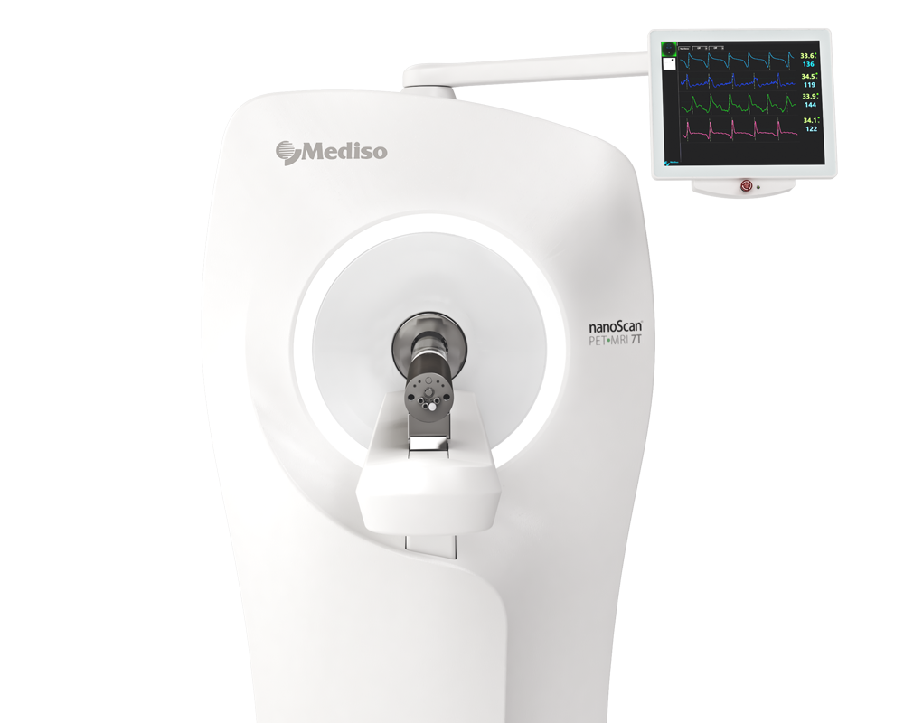
For the imaging studies, Experiments were performed on a nanoScan® PET/MRI 3T preclinical animal system. The scanner was equipped with gradients with a maximum amplitude of 550 mT/m and a maximum slew rate of 4500 T/m/s. First-order shimming was applied to improve magnetic field homogeneity before each experiment. 2D flyback EPSI sequence began with the slice selection (without water or fat suppression) followed by phase-encoding. The experiments were performed with one, two, three, or four interleaved gradient echo trains with 0.15 ms ramps of trapezoidal gradient pulses. The minimum flyback (rewind gradient) time was 0.6 ms (top time 0.3 ms and 2 × 0.15 ms ramps). We note that the amplitude and length of rewind gradient depends on the echo spacing. Ghost peaks of water and fat spectral lines were visualized with a phantom (Fig. 1) containing vegetable oil and water solution of MnCl2 (0.23 mM). The phantom’s T2 relaxation times mimics subcutaneous fat (T2 ~ 70 ms) and muscle water (T2 ~ 30 ms). The phantom experiments were performed with the maximum available FOV 80 × 80 mm, 192 phase-encoding steps, 256 sampled points per echo (matrix 192, 256), resolution in-plane 0.31 × 0.42 mm, spacing between echoes ΔTE 0.8 ms (sBW 9.74 ppm), slice thickness 2 mm, 2 averages, TR/TE1 220/3 ms, flip angle 30°, and with rBW 114 890, 217 014, 325 521, and 434 028 Hz. Readout bandwidths correspond to readout gradients (Gread) of 33.73, 63.71, 95.57, and 127.42 mT/m, respectively. It was possible to perform measurement with one echo train with ΔTE 1.55 ms (sBW 5.03 ppm) and rBW 434 028 Hz. A typical PET-MRI experiment begins with PET scans that frequently take 1–2 h. In this study, images of the rats (in vivo and post-mortem) were measured with four interleaved gradient echo trains and with four echoes in each train. Twenty-seven coronal or axial slices were measured with FOV 80 × 80 mm, matrix (192, 256), resolution in-plane 0.31 × 0.42 mm, slice thickness 1.3 mm, gap 0.2 mm, TR/TE1 420/3 ms, ΔTE 0.62 ms (sBW 12.6 ppm), flip angle 60°, 2 averages, and rBW 169 837 Hz. The acquisition time was 10 min and 53 s. Spectra of the rat were measured with two echo trains and a total of 96 echoes (1 slice, FOV 80 × 80, matrix 192 × 256, resolution in-plane, 0.31 × 0.42, 2 averages, TR/TE1 200/3, ΔTE 0.8 ms (sBW 9.74 ppm), flip angle 45°, rBW 434 028 Hz, acquisition time 2 min and 37 s).
After the phantom studies and analyzing spectrum data, water, fat, and water-fat-shift (WFS) artifact free images of the rat were measured with four interleaved echo trains and with four echoes in each train. Post-mortem and in vivo images of the rats are shown in Figs. 5. and 6, respectively. It should be noted that in vivo images (Fig. 6) were acquired from the regions that are most influenced by motions caused by the heart, lungs, and peristaltic motions of the intestines. The examples of the rat spectra measured a few minutes after its death are shown in Fig. 7.
Fig. 5:
Fig. 6.:
Fig. 7.:
- This work demonstrates the feasibility of echo-planar spectroscopic imaging with flyback readout gradients in preclinical MR systems.
- The proposed approach provides broad sBW and good water-fat signal separation for even short echo trains. Narrow rBW can be achieved by increasing the number of interleaved gradient echo trains.
- It improves SNR without the undesired consequence of WFS artifacts that are eliminated during data processing.
- Position, number, and intensity of the ghost spectral lines can be controlled by the suitable choice of sBW, number of echo train interleaves, and the number of echoes in each interleave.
W czym możemy pomóc?
Skontaktuj się z nami aby uzyskać informacje techniczne i / lub wsparcie dotyczące naszych produktów i usług.
Napisz do nas