Antiretroviral therapy timing impacts latent tuberculosis infection reactivation in a tuberculosis/simian immunodeficiency virus coinfection model
2021.12.02.
Riti Sharan et al, The Journal of Clinical Investigation., 2021
Abstract
Studies using the nonhuman primate model of M. tuberculosis /Simian Immunodeficiency Virus co-infection have revealed protective CD4+ T cell-independent immune responses that suppress LTBI reactivation. In particular, chronic immune activation rather than the mere depletion of CD4+ T cells correlates with reactivation due to SIV co-infection. Here, we administered cART at 2 weeks post-SIV co-infection to study if restoration of CD4+ T cell immunity occurred more broadly, and if this prevented reactivation of LTBI compared to cART initiated at 4 weeks post-SIV. Earlier initiation of cART enhanced survival, led to better control of viral replication and reduced immune activation in the periphery and lung vasculature thereby reducing the rate of SIV-induced reactivation. We observed robust CD8+ T effector memory responses and significantly reduced macrophage turnover in the lung tissue. However, skewed CD4+ T effector memory responses persisted and new TB lesions formed post SIV co-infection. Thus, reactivation of LTBI is governed by very early events of SIV infection. Timing of cART is critical in mitigating chronic immune activation. The novelty of these findings mainly relates to the development of a robust animal model of human Mtb/HIV co-infection that allows the testing of underlying mechanisms.
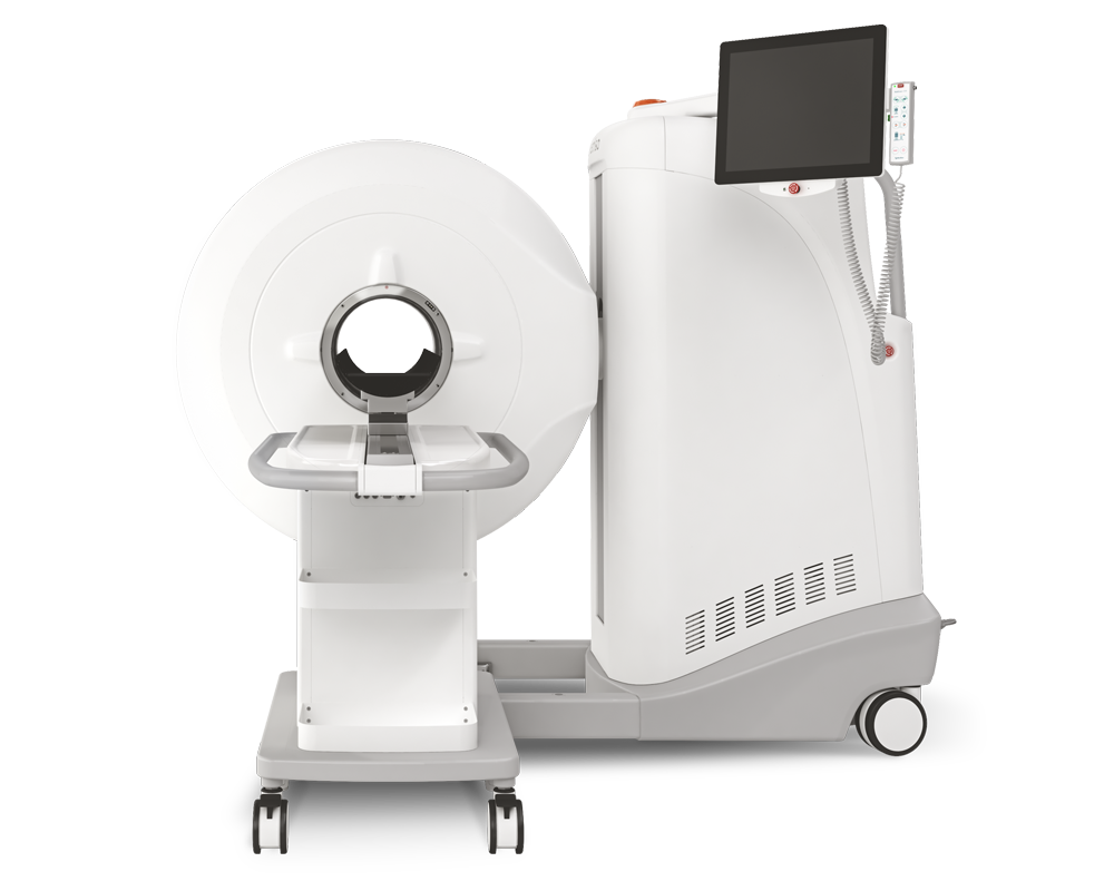
Results from MultiScan™ LFER PET/CT
Computed tomography (CT) imaging was performed on the macaques in group cART/2 week at different time points throughout the study to examine TB lesions pre-, post-SIV and post-cART. CT helped identify the lesions, TB reactivation and the granulomatous regions of the lungs. CT scans demonstrated that the worsening of the pathology was significantly mitigated in group cART/2 week, earlier cART initiation was unable to rescue from new TB lesions.
The macaques were anaesthetized (Telazol 2-6 mg/kg) and intubated to perform end-respiratory breath-hold using a remote breath-hold switch.

Fig 3. (F) Computed tomography imaging was performed on the macaques that received cART 2 weeks post-SIV co-infection at different time points throughout the study to examine the TB lesions pre-, post-SIV and post-cART

Fig 3. (G) Computed tomography imaging was performed on the macaque that received cART 4 weeks post-SIV co-infection at different time points throughout the study to examine the TB lesions pre-, post-SIV and post-cART.
Hogyan segíthetünk Önnek?
További termékinformációkért, vagy támogatásért keresse szakértőinket!
Vegye fel a kapcsolatot