Dynamic multi-pinhole collimated brain SPECT of Parkinson’s disease by [123I]FP-CIT: a feasibility study of fSPECT
2024.03.19.
Filip L. H. et al., Nature, 2024
Summary
We investigated the feasibility of using a dopamine transporter (DaT) tracer ligand ([123I]FP-CIT) along with novel multi-pinhole brain collimators for dynamic brain single photon emission computed tomography (SPECT) in suspected Parkinson's disease patients. Thirteen patients underwent dynamic tracer acquisitions before standard imaging.
Initial tests with a three-headed SPECT/CT camera (AnyScan® TRIO SPECT/CT, Mediso Medical Imaging System, Hungary) with multi-pinhole collimators, designed specifically for brain imaging, reveal a 3.9-fold increase of peak spatial sensitivity (700 cps/MBq) and a spatial resolution of approximately 5 mm half that of conventional two-headed SPECT with parallel-hole collimators. The improved resolution lowers partial volume effects (PVE) and improves delineation of striatum while the higher sensitivity enables dynamic scanning and time framing with the potential to more accurately reveal tracer dynamics and hence the pharmacokinetics of [123I]FP-CIT.
Uptake values were corrected for partial volume effects. Specific binding ratio (SBRcalc) was calculated, reflecting binding potential relative to non-displaceable binding (BPND) in the cortex. Additional pharmacokinetic parameters (BPND, R1, k2) were estimated using the simplified reference tissue model, revealing differences between Kahraman low-score (LS) and high-score (HS) groups.

Volumes of interest as shown on a high-score patient scan. Pink: Vcaudate nucleus. Red: Vputamen. VS = Vcaudate nucleus + Vputamen. White: Vmargin. Blue (rectangularly shaped): VBG. Green: Occipital cortex. Orange: Cerebellum.
Results showed increasing striatal tracer uptake until 100 min post-injection, with consistent values afterward.

Relation between activity and time. (a) Uncorrected mean activity concentration for striatum. (b) Striatal mean activity concentration corrected for partial volume and minor movement artefacts. (c) Corrected mean activity concentration in the caudate nucleus. (d) Corrected mean activity concentration in the putamen. (e) Mean activity concentration in the occipital cortex. (f) Mean activity concentration in the cerebellum. Each different marker represents a patient with values from both the left and right striatum shown in (a–d). Patients #1–4 had Karahman scores of < 4 at 3-h images and are illustrated in orange.

Calculated specific binding ratio (SBRcalc) for all patients throughout the scan duration. Each patient is represented by a different mark and illustrated with a dataset from both the left and the right striatum. Patients with #1–4 had Karahman scores of < 4 at 3 h p.i. images and are illustrated in orange.
Uptake and SBRcalc ratios matched visual assessment. LS patients had lower putamen than caudate nucleus tracer uptake, decreased BPND values, while R1 and k2 values were comparable to HS patients. In conclusion, dynamic multi-pinhole SPECT using DaT tracer with the extraction of pharmacokinetic parameters is feasible and could help enable early differentiation of reduced and normal DaT values.
Full article on Nature
-
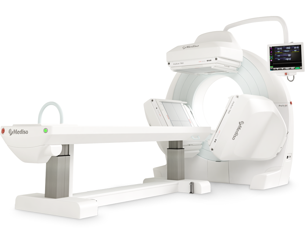
AnyScan® TRIO SPECT
Ultra-fast Triple-NaI-Detector SPECT for wide range of clinical applications
-
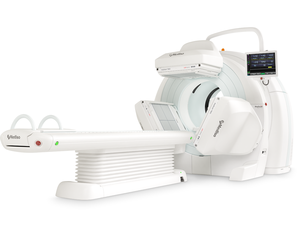
AnyScan® TRIO SPECT/CT
Ultra-fast Triple-NaI-Detector SPECT system for wide range of clinical applicati...
-
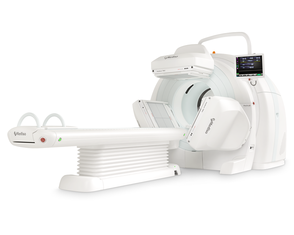
AnyScan® TRIO SPECT/CT/PET
An integrated triple-detector SPECT/CT and PET/CT system in a single-room instal...
-
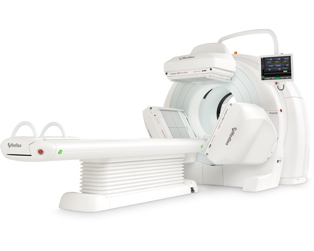
AnyScan® TRIO-TheraMAX SPECT/CT
Jakość Obrazów Teranostycznych i Diagnostycznych na MAXymalnym poziomie


W czym możemy pomóc?
Skontaktuj się z nami aby uzyskać informacje techniczne i / lub wsparcie dotyczące naszych produktów i usług.
Napisz do nas