AI-based synthetic CT attenuation correction enables reliable quantitative SPECT in unilateral condylar hyperplasia
2026.04.20.
Kovács A.R. et al., EJNMMI Phys, 2026
Summary
Unilateral condylar hyperplasia (UCH) is a rare mandibular growth disorder characterized by progressive enlargement of one condylar process. Bone SPECT/CT using 99mTechnetium labeled diphosphonates are widely used for its evaluation. CT-based attenuation correction (CTAC) increases radiation exposure and may be affected by registration errors. As a CT-independent, non-irradiating alternative, artificial intelligence (AI)-generated synthetic CT (SyCT) has been proposed, which aligns with the values of the ALARA principle. Networks like convolutional neural network (CNN) or generative adversarial network (GAN) can create CT-equivalent attenuation maps directly from emission data without requiring a CT scan.
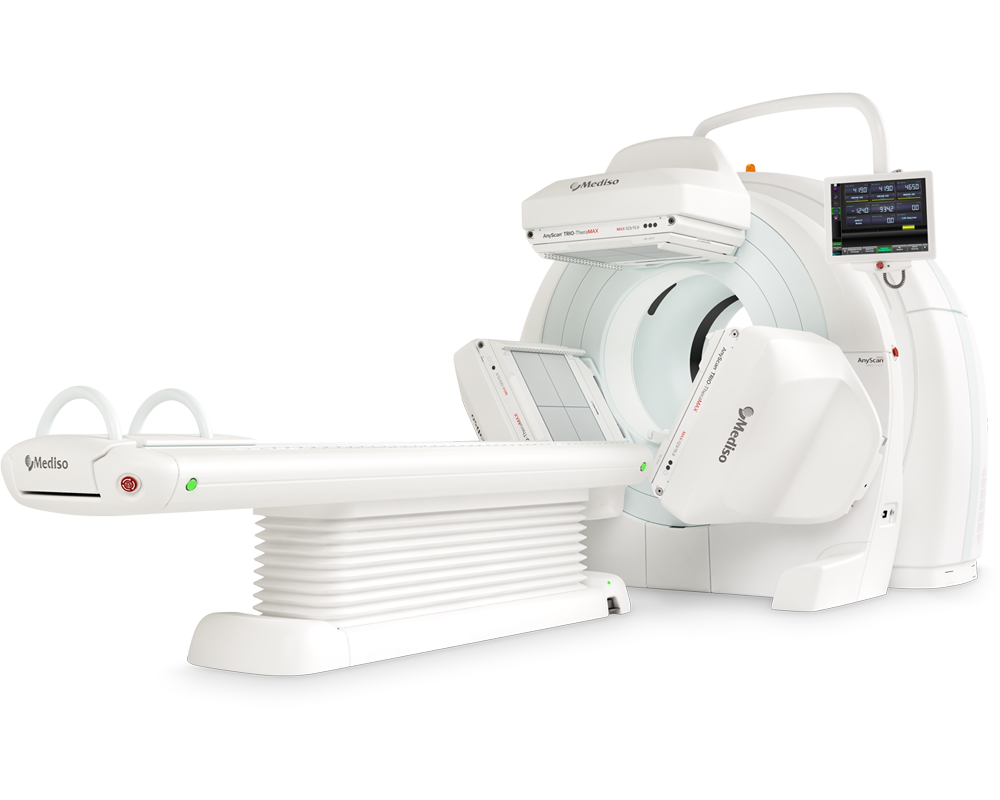
AnyScan® TRIO SPECT/CT (Mediso Ltd., Budapest, Hungary) with low energy high resolution high sensitivity (LEHR-HS) collimators were used for the SPECT imaging and CT images were acquired in the same equipment without contrast enhancement.
SPECT reconstructions were then performed using both the original CT and the SyCT, applying identical parameters in Interview XP version 3.09 (Mediso Ltd., Budapest, Hungary). The SPECT images reconstructed with the original CT were designated as CTAC (CT Attenuation Correction), while those reconstructed using the AI- generated SyCT were designated as SyCTAC (Synthetic CT Attenuation Correction). The image processing was performed using the Interview Fusion 3.10.009.0000 reporting and evaluation software (Mediso Ltd., Budapest, Hungary).
During the evaluation, the right and left mandibular condylar regions were identified on the CT-s, spherical VOIs with a diameter of 2 cm were placed over both condyles on the CTAC images. Using the same approach, a 2 cm spherical VOI was also placed over the clivus region. Subsequently, the condylar and clivus VOIs were copied to the SyCTAC images in the identical positions. Based on the VOIs placed over the mandibular condyles and the clivus, SUVmax and SUVmean values were determined on both the CTAC and SyCTAC images and two kinds of uptake values (relative uptake fractions and relative uptake normalized to the clivus as reference area) were calculated.
Statistical analyses were performed using SPSS version 31 (IBM Corp., Armonk, NY, USA). Normality was assessed with the Shapiro-Wilk test. Agreement between the two quantitative approaches (CTAC-based vs. SyCTAC- based ratios) was evaluated differently for the two groups of uptake indices. Bland-Altman analysis was applied to the fractional uptakes of the “affected” side. For the uptake values normalized to a reference area, the measurements from the two sides are not statistically independent. To assess agreement between the two methods, we used a mixed-effects model with 'patient' as a random effect, thereby accounting for the correlation between the two condyles belonging to the same individual.
Results from TheraMAX SPECT/CT
Visual evaluations were independently performed by three nuclear medicine specialists, who reported no substantial differences between the SPECT images reconstructed with CT and those reconstructed with SyCT. Specifically, no differences were noted regarding image intensity display, lesion detectability, or the feasibility of VOI placement.

Axial, coronal and sagittal slices of CT (A) and SyCT (B) images.
Axial slices of the CTAC (C) and SyCTAC (D) images with spherical VOIs (diameter: 2 cm) placed over both condyles and the clivus.
Quantitative evaluation and statistical analysis (such as tests of normality, Bland-Altman analysis of the relative uptake fractions, Analysis of the relative uptake normalized to the clivus and differences between SyCTAC) was made and CTAC-based relative uptake values were examined as well.
According to the Shapiro-Wilk tests, the distributions of the calculated relative uptake ratios derived from CTAC and SyCTAC images did not differ significantly from a normal distribution and the Blant-Altman analysis also shows that the possible differences are clinically not significant.
This study examined whether AI-generated SyCT images can provide attenuation maps suitable for quantitative 99mTc-MDP bone SPECT reconstruction in patients with UCH. The visual assessment performed by three nuclear medicine specialists confirmed that SyCTAC-based reconstructions did not differ perceptibly from CTAC in overall image quality, lesion visibility, or suitability for VOI placement. Across the evaluated parameters, SyCTAC demonstrated close agreement with CTAC in the quantification of condylar uptake, when uptake values were normalized to the summed activity of both condyles, although tended to slightly underestimate the update fraction of the affected side.
Bland-Altman analysis confirmed no evidence of clinically significant bias or proportional error. Greater variability was observed in ratios normalized to the clivus, with statistically significant differences between SyCTAC and CTAC. These discrepancies may relate to the anatomical and functional characteristics of the clivus, a dense osseous structure with relatively low metabolic activity, making SUV estimation more sensitive to small deviations in attenuation mapping.
Nonetheless, the significant correlations observed for both maximum and mean clivus-normalized ratios indicate that SyCTAC still preserved the overall ranking of uptake values. Importantly, clinical assessment of UCH most commonly relies primarily on condyle-to-condyle comparisons rather than reference-region normalization, reducing the practical impact of these differences.
W czym możemy pomóc?
Skontaktuj się z nami aby uzyskać informacje techniczne i / lub wsparcie dotyczące naszych produktów i usług.
Napisz do nas