Performance evaluation of a novel multi‑pinhole collimator on triple‑NaI‑detector SPECT/CT for dedicated myocardial imaging
2023.03.25.
Krizsan et al., EJNMMI Physics, 2023
Background
In this study we evaluated the imaging capabilities of a novel Multipinhole collimator (MPH-Cardiac) specially designed for nuclear cardiology imaging on a Triple-NaI-detector based SPECT/CT system.
Methods
99mTc point source measurements covering the field of view (FOV) were used to determine tomographic sensitivity ( TSpointsource) and spatial resolution. Organsize tomographic sensitivity ( TSorgan) was measured with a left ventricle (LV) phantom filled with typical myocardial activity of a patient scan. Reconstructed image uniformity was measured with a 140 mm diameter uniform cylinder phantom. Using the LV phantom once filled with 99mTc and after with 123I, Contrast-to-noise ratio (CNR) was measured on the reconstructed images by ROI analysis on the myocardium activity and on the LV cavity. Furthermore, a polar map analysis was performed determining Spill-Over-Ratio in water ( SORwater) and image noise. The results were compared with that of a dual-head parallel-hole low energy high resolution (LEHR) collimator system. A patient with suspected coronary artery disease (CAD) was scanned on the LEHR system using local protocol of 16 min total acquisition time, followed by a 4-min MPH-Cardiac scan.
Triple‑NaI‑detector SPECT system with multi‑pinhole collimator

C-shape detector configuration of the triple-NaI-detector SPECT/CT system with MPH-Cardiac collimators (A); Schematic figure of projection trajectories of myocardium activity distribution for the MPH-Cardiac collimator system (B); A photograph of the rear view of the MPH-Cardiac collimator (C); Single projection image of the anthropomorphic phantom LV insert filled with 99mTc (D)
Tomographic sensitivity
Peak tomographic sensitivity measured with the point source was found to be 1013 cps/ MBq in the absolute axial center of the FOV (CFOV) while it is decreasing toward the radial edges: 951 cps/MBq at 5 cm radial offset from the CFOV and 641 cps/MBq at 10 cm radial from the CFOV.

Axial (A) and Radial (B) tomographic sensitivity profiles of the MPH-Cardiac collimator system. The axial sensitivity profile was measured at 50 mm and 100 mm radial offsets. The reference line of the LEHR tomographic sensitivity is displayed as a dashed purple line.

LV size tomographic count sensitivity (TSorgan) data measured on reconstructed images of the DataSpectrum LV phantom in case of AnyScan SC LEHR and AnyScan TRIO MPH-Cardiac, compared to several cardiac SPECT imaging systems of different vendors.
Spaital resolution
The absolute average spatial resolution was found to be 4.38 mm FWHM calculated from the measurements with the MPH-Cardiac collimators along the central axis of the FOV and through the centrum of the FOV in the radial directions. Along with the St.Dev. values. Spatial resolution results are presented using the dedicated reconstruction method (Tera-Tomo™ 3D SPECT-Q).

Mean and Standard Deviation (St. Dev.) values of FWHM spatial resolution results calculated on the reconstructed images, measured along the central axis and through the centrum of the FOV in the radial directions.
Image uniformity
Reconstructed SPECT images of the uniformity cylinder measurements. The VOI analysis resulted 0.292% versus 0.214% uniformity values for the LEHR and MPH-Cardiac measurements, respectively.

Schematic figure and real images of the uniform cylinder (140 mm diameter) filled with 99mTc acquired on the AnyScan SC LEHR system and AnyScan TRIO with MPH-Cardiac collimator system. The upper limit of the inverted gray color bar compared to the maximum intensity in percentage is also indicated.
CNR, Spill‑over‑ratio and noise analysis
CNR was found to be 15.5 in case of MPH-Cardiac 11.7 for LEHR in case of 99mTc, while it was 13.5 and 8.3, respectively, when using 123I. Polar map analysis was performed on the LV phantom measurements with 99mTc and 123I. The MPH-Cardiac images showed much better contrast and image noise compared to the conventional LEHR technique as can be depicted in Fig. 4 and Table 3. Perfusion polar maps revealed SORwater values of 28.8% versus 21.1% for the 99mTc measurements with the LEHR and MPH-Cardiac configuration, respectively, as indicated in Table 3. In case of the 123I measurements, SORwater values were found to be 31.4% versus 24.3% for the LEHR and MPH-Cardiac measurements, respectively. Pixel noise of the 99mTc polar maps resulted values of 0.38% versus 0.24% for the LEHR and MPH-Cardiac, respectively. Meanwhile, 123I polar maps resulted values of 0.62% versus 0.21% for the LEHR and MPH-Cardiac, respectively. Visually interpreting the polar maps, in each case the MPH-Cardiac measurements resulted in better image contrast compared to the LEHR measurements for both isotopes despite of the shortened acquisition time as can be seen in figure.

Polar Map representations of the Data Spectrum LV insert phantom filled with 99mTc (left upper row) and 123I (right upper row) measured with the AnyScan SC with LEHR 16 min acquisition and the AnyScan TRIO with MPH-Cardiac collimator 4 min acquisition. Short axis images of the reconstructed LV inserts are presented for the 99mTc (left lower row) and 123I (right lower row) measurements. Half-moon shaped ROI was applied on both the myocardium region of the phantom (green) and a circular ROI within the LV cavity (yellow). These ROIs were applied on all four LV SPECT images.
Patient scan
Reconstructed images and perfusion polar maps for the 16 min LEHR acquisition and 4 min MPH-Cardiac acquisition of the same patient are presented in figure. The reconstructed images revealed better contrast for MPH-Cardiac, as well as more homogenous polar map in the anterior wall region.

Representative post-stress SPECT image slices and polar maps of a 67 years old male CAD patient. Reconstructed images of the four minutes MPH-Cardiac acquisition (upper row) and sixteen minutes LEHR acquisition (lower row).
Conclusions
Significant image quality improvement can be achieved with a novel multi-pinhole technology, the MPH-Cardiac combined with triple-NaI-detector SPECT, when comparing to conventional parallel-hole LEHR collimator dual-detector SPECT. This improvement can be attributed to the increased tomographic sensitivity and uncompromised spatial resolution in the myocardium region, provided by the novel collimator design. Image quality improvement in MPH-Cardiac with triple-NaI-detector SPECT paves the way for shorter acquisition times and lower effective doses for both perfusion and innervation imaging applications in nuclear cardiology.
Full article on EJNMMI Physics.
-
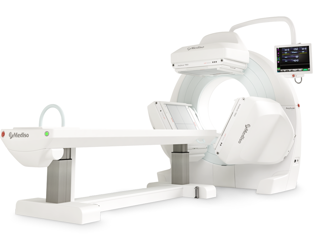
AnyScan® TRIO SPECT
Ultra-fast Triple-NaI-Detector SPECT for wide range of clinical applications
-
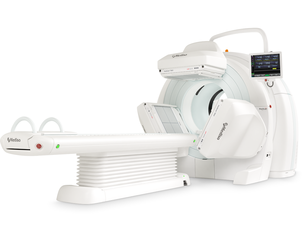
AnyScan® TRIO SPECT/CT
Ultra-fast Triple-NaI-Detector SPECT system for wide range of clinical applicati...
-
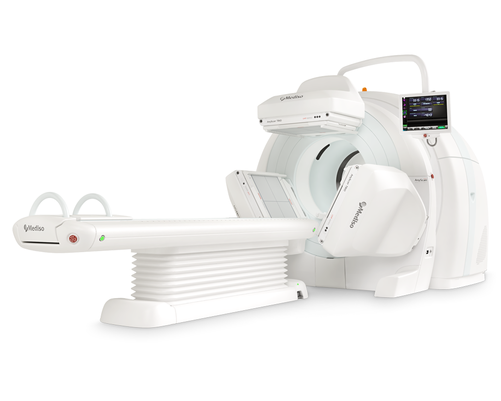
AnyScan® TRIO SPECT/CT/PET
An integrated triple-detector SPECT/CT and PET/CT system in a single-room instal...
-
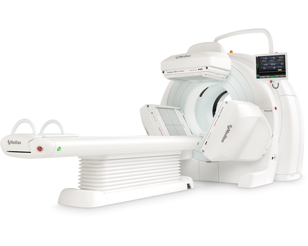
AnyScan® TRIO-TheraMAX SPECT/CT
Theranostic and Diagnostic Imaging with MAXimum Performance


Hogyan segíthetünk Önnek?
További termékinformációkért, vagy támogatásért keresse szakértőinket!
Vegye fel a kapcsolatot