Summary
Alzheimer's disease (AD) is the most common form of dementia with an estimated number of more than 40 Million cases worldwide. To date, a final diagnosis of the disease can only be made by histopathological detection of amyloid plaques and neurofibrillary tangles post mortem, therefore it is mandator to find proper animal models, on which different in vivo diagnostics can be used.
The imaging biomarkers of AD that are able to detect molecular changes in vivo and transgenic animal models mimicking AD pathologies are essential for the evaluation of new therapeutic strategies. Positron-emission tomography (PET) using either 18F-Fluorodeoxyglucose (18F-FDG) or amyloid-tracers is a well-established, non-invasive tool in the clinical diagnostics of AD assessing two major pathological hallmarks. 18F-FDG-PET is able to detect early changes in cerebral glucose metabolism and amyloid-PET shows cerebral amyloid load. However, the suitability of 18F-FDG- and amyloid-PET in the widely used 5XFAD mouse model of AD is unclear as only a few studies on the use of PET biomarkers are available showing some conflicting results. The aim of this study was the evaluation of 18F-FDG-PET and amyloid-PET in 5XFAD mice in comparison to neurological deficits and neuropathological changes.
Results from the nanoScan PET/MRI
For the small animal imaging, the authors have used a nanoScan PET/MRI 1T, which provided a good enough spatial resolution to differentiate the examined brain regions, and creating even 10mm3 large VOIs (which is still above the nanoScan PET intrinsic resolution of 0.7mm).
18F-FDG-PET/MRI was performed on 7- and 12-month-old male 5XFAD mice as well as age- and sex-matched C57Bl/6J wild type mice (n = 4–6 per group). 18F-FDG (11.46–20.53 MBq; mean 16.81 MBq) was injected into a tail vein with a maximum volume of 200 μl followed by an uptake period of 45 min. Mice were awake during the uptake process. PET scans were performed for 20 min. MRI-based attenuation correction was conducted with the material map (matrix 144 × 144 × 163 with a voxel size of 0.5 × 0.5 × 0.6 mm3, repetition time: 15 ms, echo time: 2.032 ms and a flip angle of 25°) and the PET images were reconstructed using the following parameters: matrix 136 × 131 × 315, voxel size 0.23 × 0.3 × 0.3 mm3.
For the 18F-FBB-PET/MRI imaging, it was performed on 7- and 12-month-old male 5XFAD mice as well as age- and sex-matched C57Bl/6J wild type mice after 18F-FDG-PET imaging (n = 4–6 per group). In isoflurane anesthetized mice 18F-Florbetaben (7.5–24 MBq; mean 14 MBq) was administered intravenously with a maximum volume of 200 μl. PET acquisition of 30 min duration started after an uptake period of 40 min.
Figure 2. shows the main results from 18F-FDG-PET images in coronal, transverse, and sagittal view. (A–C) MRI images with predefined brain regions. (D–F) 18F-FDG images of a representative 7-month-old WT mouse. (G–I) 18F-FDG-PET images of a 7-month-old 5XFAD mouse. 7-month-old 5XFAD mice showed distinctly lower FDG uptake compared to WT mice. (J–L) 18F-FDG images of a 12-month-old WT mouse. 12-month-old 5XFAD mice also showed significantly lower 18F-FDG uptake compared to age-matched WT mice. A, Amygdala; Bs, Brain Stem; C, Cortex; Cb, Cerebellum, H, Hypothalamus; Hc, Hippocampus; Hg, Harderian Glands; M, Midbrain; O, Olfactory Bulb; S, Septum/Basal Forebrain; St, Striatum; T, Thalamus.
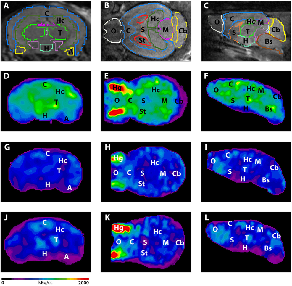
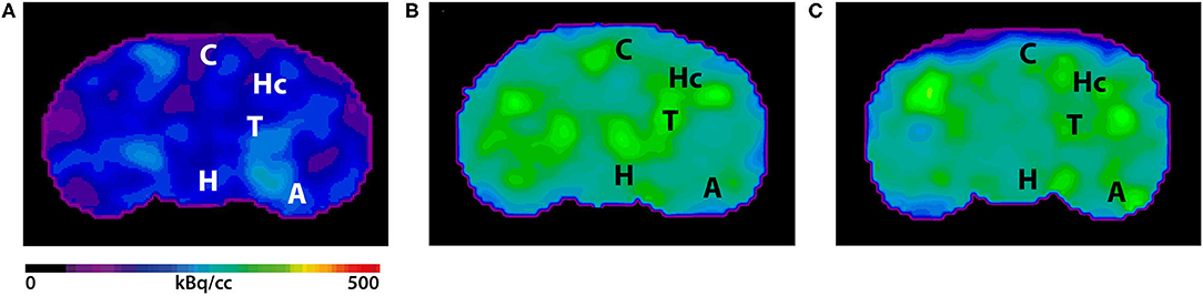
Figure 4. shows the results from 18F-Florbetaben-PET images in coronal view. (A) 18F-Florbetaben image of a 7-month-old WT mouse. (B) Cerebral 18F-Florbetaben uptake was higher in 7-month-old 5XFAD mice as well as in 12-month-old 5XFAD mice (C).
- The authors could show that PET biomarkers 18F-FDG and 18F-Florbetaben detected cerebral hypometabolism and increased plaque load even before the onset of severe memory deficits.
- Summarizing their results, the 18F-FDG- and 18F-Florbetaben-PET, are useful tools for the in vivo detection of cerebral AD pathologies in 5XFAD mice even before the onset of severe memory deficits. The 5XFAD mouse model of AD therefore is a suitable model for preclinical PET studies showing comparable changes to AD patients with the clinically established biomarkers 18F-FDG and 18F-Florbetaben. Therefore, PET imaging can be utilized as a readout for therapeutic effects in vivo in future longitudinal therapy studies using the 5XFAD mouse model of AD.
Full article on frontiersin.org


