Temperature-Dependent Modulation of Cardiac Metabolism, Post-Injury Survival and Regenerative Rate in Axolotls
2026.07.13.
Anita Dittrich et al., Metabolites, 2026
Abstract
Background/Objectives: Cardiac regenerative ability varies in vertebrates. Adult mammals cannot mount a regenerative response, while fetal mammals and some salamanders and teleosts fully regenerate the heart after a cryoinjury mimicking a myocardial infarction. This contrast is suggested to be regulated in part by metabolism, with high regenerative capacity correlating with a comparatively lower mass-specific metabolic rate, ectothermy rather than endothermy and a metabolic phenotype favoring glycolysis in cardiac muscle. Methods: In this physiological study on axolotl salamanders, we altered the housing temperatures from the standard 20 °C to 10 °C, 25 °C and 30 °C and assayed key metabolic parameters as well as cardiac function, survival and regenerative capacity. Results: Our study demonstrated that while axolotls could be housed at temperatures ranging from 10 °C to 30 °C in an uninjured state, signs of a pathological response involving cardiac and metabolic insufficiency and mortality, especially after cryoinjury, increased progressively with increasing temperatures. We observed several metabolic effects, including differences in oxygen consumption, plasma metabolites and cardiac function. Cardiac regeneration after cryoinjury progressed as expected with only a small remaining injury after 60 days at the standard housing temperature of 20 °C. Regeneration was highly reduced in a reversible manner at 10 °C while regenerative rate was not affected at 25 °C. At 30 °C, cardiac regeneration could not be evaluated as the majority of animals (five out of six) did not survive the injury, likely reflecting insufficient cardiac reserve capacity to simultaneously sustain thermal metabolic effects and support tissue repair. Conclusions: The ectothermic axolotl undergoes several metabolic changes when exposed to different housing temperatures, with heart regeneration showing a narrower permissive temperature range than survival of the axolotl in an uninjured state.
Results from nanoScan® PET/MRI
Uninjured and anesthetized animals were injected with [18F]-FDG (~25 MBq/animal) intravenously in the jugular vein and allowed 2 h of circulation time at their set housing temperature (with or without aeration). [18F]-FDG is a glucose analog that cannot be metabolized by the cells, and thus, accumulation of FDG is a proxy for glucose uptake. The appropriate circulation time for FDG to reach steady-state for PET imaging has been previously determined in axolotls. Combined PET and MRI imaging was obtained on a Mediso NanoScan PET/MRI system (Mediso, Budapest, Hungary) to detect [18F] decay with an isotropic image resolution of 0.4 mm and an acquisition time of 20 min for PET and an additional 20 min for MRI. The axolotls were wrapped in moist paper towels soaked in anesthetic fluid throughout the 40 min scan and immediately returned to housing water afterwards. Signal values were measured within regions of interest in the different tissues using ImageJ (version 1.54a) and reported as values relative to the signal in the brain.
Figure 4. Metabolic effects of acute exposure to different temperatures in uninjured axolotls in vivo. (A) Oxygen consumption assayed by closed respirometry. (B) Blood glucose and (C) ketones measured with glucometer. (D) Blood radioactivity after 2 h of circulation with [18F]-FDG. (E) [18F]-FDG uptake in different tissues relative to the brain in pilot animals in aerated water anesthetized with benzocaine. (F) [18F]-FDG uptake in different tissues relative to the brain in animals anesthetized with propofol in aerated water. (G) [18F]-FDG uptake in different tissues relative to the brain in animals anesthetized with propofol in non-aerated water. (H) Coronal (top row) and sagittal (bottom row) maximum intensity projections of positron emission tomography images of [18F]-FDG (glucose analog) distribution after 2 h circulation time with propofol anesthesia at 10 °C, 20 °C, 25 °C, and 30 °C in aerated and non-aerated conditions. Animals used for blood sampling (B,C) were on the same day used for PET imaging under propofol anesthesia (F) with n = 6 for each temperature. The same batch of animals was used for the experiments shown in (E,G) but with ample time in between experiments in order to completely wash out anesthesia and return to standard temperature. Error bars represent standard deviation. Asterisk indicates significant differences: * = p < 0.05, ** = p < 0.005 and *** = p < 0.005. In the case of O2 consumption rate, multiple comparison of all other groups compared to the 20 °C control was done by one-way ANOVA with Dunnett’s post hoc tests. For glucose, ketones and blood radioactivity, the same tests were done instead with Brown–Forsythe and Welch ANOVA due to unequal standard deviations among the groups. For PET imaging tests were done with one-way or two-way ANOVA (depending on whether different temperatures were involved) with Tukey post hoc tests comparing all tissues and temperatures when applicable.
 Figure 4. Metabolic effects of acute exposure to different temperatures in uninjured axolotls in vivo. (A) Oxygen consumption assayed by closed respirometry. (B) Blood glucose and (C) ketones measured with glucometer. (D) Blood radioactivity after 2 h of circulation with [18F]-FDG. (E) [18F]-FDG uptake in different tissues relative to the brain in pilot animals in aerated water anesthetized with benzocaine. (F) [18F]-FDG uptake in different tissues relative to the brain in animals anesthetized with propofol in aerated water. (G) [18F]-FDG uptake in different tissues relative to the brain in animals anesthetized with propofol in non-aerated water. (H) Coronal (top row) and sagittal (bottom row) maximum intensity projections of positron emission tomography images of [18F]-FDG (glucose analog) distribution after 2 h circulation time with propofol anesthesia at 10 °C, 20 °C, 25 °C, and 30 °C in aerated and non-aerated conditions. Animals used for blood sampling (B,C) were on the same day used for PET imaging under propofol anesthesia (F) with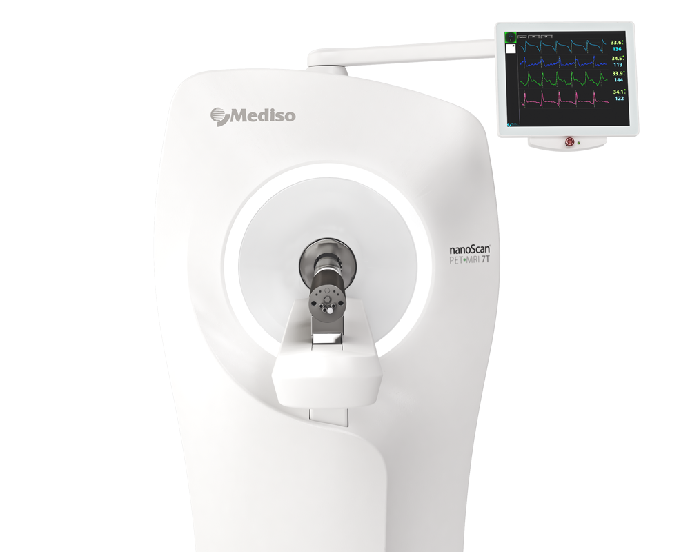
¿como podemos ayudarlo?
Póngase en contacto con nosotros para obtener información técnica, productos y servicios!
Ponerse en contacto