18F-FDG PET/MRI Imaging in a Preclinical Rat Model of Cardiorenal Syndrome—An Exploratory Study
2022.12.06.
Dan Mihai Furcea et al. International Journal of Molecular Science, 2022
Summary
Cardiorenal syndrome (CRS) is an intricate clinical condition where chronic kidney disease (CKD) and heart failure (HF) coexist and evolve by system crosstalk, and the failure of one organ accelerates the progression of structural damage and failure in the other organ. When the acute or chronic malfunction of the left ventricle co-evolving with acute or chronic renal disease, the emerging clinicopathological entity is classified as CRS, and it leads to the progressive deterioration of both systems resulting in high morbidity and mortality.
CRS denotes the bidirectional interaction of chronic kidney disease and heart failure with an adverse prognosis but with a limited understanding of its pathogenesis. The aim was to investigate the correlates between biochemical blood markers, histopathological and immunohistochemistry features, and 2-deoxy-2-fluoro-D-glucose positron emission tomography (18F-FDG PET) metabolic data in low-dose doxorubicin-induced heart failure, cardiorenal syndrome, and renocardiac syndrome induced on Wistar male rats. This is the first study that investigates the underlying mechanisms for CRS progression in rats using 18F-FDG PET. Clinical, metabolic cage monitoring, biochemistry, histopathology, and immunohistochemistry combined with PET/MRI (magnetic resonance imaging) data acquisition at distinct points in the disease progression were employed for this study in order to elucidate the available evidence of organ crosstalk between the heart and kidneys. In used CRS model, was found that chronic treatment with low-dose doxorubicin followed by acute 5/6 nephrectomy incurred the highest mortality among the study groups, while the model for renocardiac syndrome resulted in moderate-to-high mortality. 18F-FDG PET imaging evidenced the doxorubicin cardiotoxicity with vascular alterations, normal kidney development damage, and impaired function. Given the fact that standard clinical markers were insensitive to early renal injury, it was believed that the decreasing values of the 18F-FDG PET-derived renal marker across the groups and, compared with their age-matched controls, along with the uniform distribution seen in healthy developing rats, could have a potential diagnostic and prognostic yield in cardiorenal syndrome.
Results from the nanoScan PET/MRI
A total of 34 Wistar albino male rats (2 months old at arrival) were divided into 4 groups: 8 animals in the control (L1), 8 in doxorubicin-induced heart failure (L2), 8 in cardiorenal syndrome (L3) and 10 in renocardiac syndrome (L4). On top of that, 4 rats were kept alive as the age control of the 18F-FDG PET imaging data, annotated as L2Aco for L2 and L3&L4Aco for L3 and L4. They were housed in individually ventilated cages with a 2-week period of acclimatization. Cardiac dysfunction was induced by the intraperitoneal injection of 1 mg/kg of doxorubicin twice a week for 6 weeks, renal failure was induced by the 5/6 nephrectomy protocol, following the guidelines. Then a deep dissection for the kidney pedicle was performed. Left posterior and inferior kidney arteries were ligated and excision of the right kidney was made en bloc. The closure of the abdominal wall was attained after the control of hemostasis and cavity wash with 3 mL of serum.
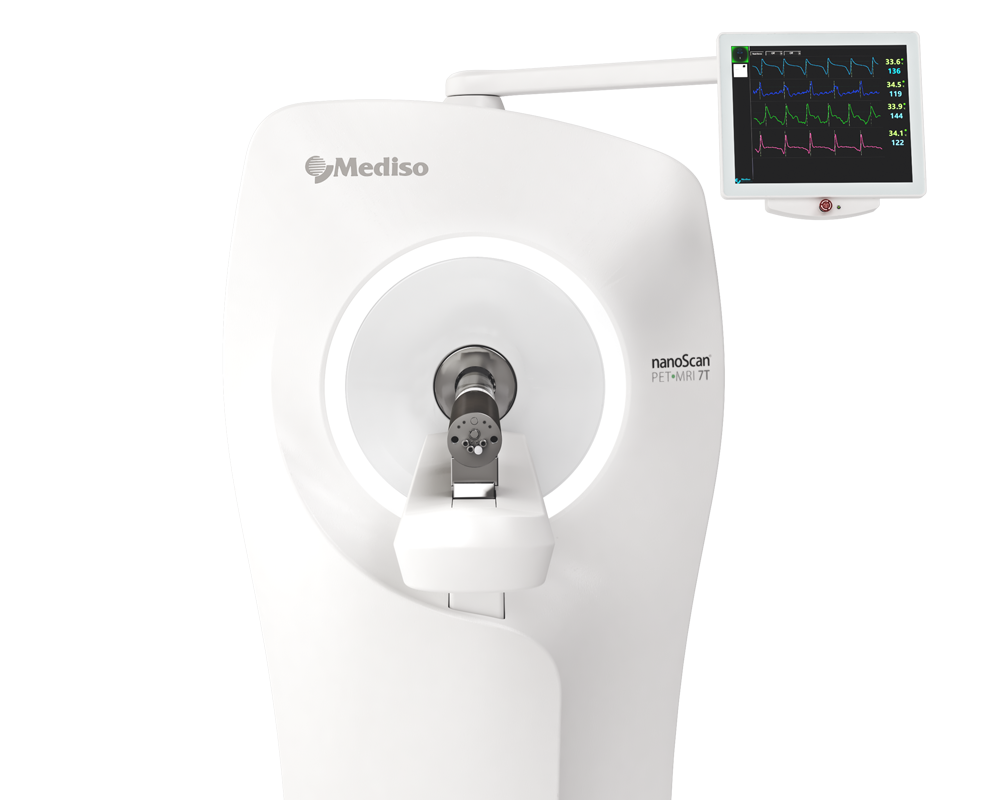
The PET/MRI acquisition was performed using nanoScan PET/MRI (Mediso®, Budapest, Hungary).
A fast MRI scout for system calibration and subject localization was performed. It was followed by an axial T1-weighted 3D spoiled gradient recalled echo sequence (GRE) for anatomical.
18F-FDG was administered through a 26G cannula (average dose of 9.56 0.7 MBq/100 g). Pre-scan glycaemia levels were measured with a clinical-grade glucometer before 18F-FDG administrations.
PET data acquisition consisted of a 40 min dynamic scan with a single FOV over the thorax and abdominal region without cardiac gating. Data acquisition was in list mode, 3D, with a 1 to 5 in-plane coincidence detection activated and a normal photon count rate.
Data reconstruction employed the proprietary Tera-Tomo engine (Mediso©) with a 3D maximum a posteriori algorithm (8 iterations and 3 subsets) using an energy window of 400–600 keV, a 0.6 mm isotropic voxel size, a small regularization term (alpha = 0.0001 and beta = e-3, and a variance-reduced delayed window for random correction. List-mode data were partitioned into a dynamic study for the first 5 min with a high-frequency sampling of 1 s, the next 15 min with 10 s per frame, and the last 20 min with 30 s per frame. Additionally, 3 static reconstructions with a 5 min interval each were performed for the standard uptake value (SUV) analysis for 0–5 (TP1), 15–20 (TP2), and 35–40 (TP3) minute periods.
- Regarding target organ metabolic activity, myocardium uptake was similar for the L1 and L2 groups throughout these intervals, while the L3 group had an almost 2-fold increase and L4 had a 1.5-fold increase compared to L1 at the last sampled interval (Figure 7 for TP2 and TP3).

Figure 7. Heart and liver close-ups in parametric maximal intensity projection images with voxel-wise normalization to blood SUV, depicting different behaviours of 18F-FDG according to time interval and group.
- Renal cortical metabolic activity manifested a peculiar behaviour, with a first-phase decrease in the distribution of radiotracer in L4 individuals compared to all other groups, followed by a marked elevation in the intermediary phase for L3 and L4 (Figure 8 for TP1 and TP2). By the time of the uptake-dominated phase, these alterations were reversed to the level of the L1 and L2 groups (Figure 8 for TP3).

Figure 8. Renal close-ups in parametric maximal intensity projection images with voxel-wise normalization to blood SUV, depicting different behaviours of 18F-FDG according to time interval and group.
¿como podemos ayudarlo?
Póngase en contacto con nosotros para obtener información técnica, productos y servicios!
Ponerse en contacto