Vascular and Neural Transcriptomics Reveal Stage-Dependent Pathways to Inflammation and Cognitive Dysfunction in a Rat Model of Hypertension
2025.08.05.
Philipp Arndt et al., Journal of the American Heart Association, 2025
Summary
Chronic arterial hypertension causes cerebral microvascular dysfunction and increases dementia risk in aging. However, cognitive health preservation by therapeutic blood pressure lowering alone is limited and depends on disease duration, the degree of irreversible tissue damage, and whether microvascular function can be restored. This study aimed to understand molecular and cellular temporospatial mechanisms of disease in the course of hypertension.
The effects of initial, early chronic and late chronic hypertension in the frontal brain of spontaneously hypertensive stroke-prone rats were investigated by applying behavioral tests, histopathology, immunofluorescence, fluorescence-activated cell sorting, microvascular/neural tissue RNA sequencing, and 18F-fluorodeoxyglucose PET imaging.
Results from nanoScan® PET/MRI
SHRSPs (non-transgenic spontaneously hypertensive stroke-prone) rats and Wistar rats in three age groups (6-8 weeks, 24-25 weeks, 32-34 weeks) were injected intraperitoneally with 18F-fluorodeoxyglucose. Animals were injected in the awake state. Positron emission tomography (PET) scanning in a small-animal PET/MRI (nanoScan, Mediso, Budapest, Hungary) started 45 minutes after injection. Static PET scans of the head were performed followed by T1-weighted gradient echo MRI for anatomic correlations and attenuation correction. List mode data from PET scans were reconstructed by Ordered Subset Expectation Maximization with 4 iterations, 6 subsets, and a voxel size of 0.4 mm isotropic.
Gene expression alterations in the hypertensive rat brain suggested an upregulation of energy-demanding processes. Using 18F-fluorodeoxyglucose PET imaging, which measures brain glucose uptake, it was assessed whether these molecular and cellular changes affect brain metabolism and are transferable to further brain regions. A standard protocol of 18F-fluorodeoxyglucose PET imaging based on a 45-minute uptake phase was used, followed by read-outs of the distribution of the trapped, phosphorylated tracer in one static scan. Increases of up to 30% in early chronic hypertensive animals compared with age-matched controls were noted. The cingulate and retrosplenial cortices as well as motor and somatosensory cortices were mostly affected (Figure 7). In late chronic hypertension, the spatial distribution of affected regions is largely similar but the effect size is markedly reduced as compared with early chronic hypertension.
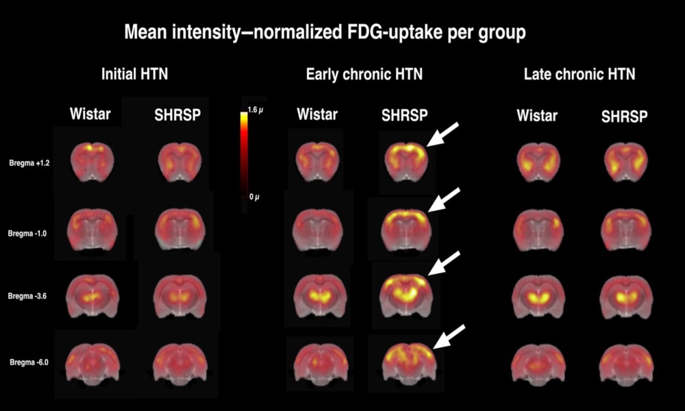
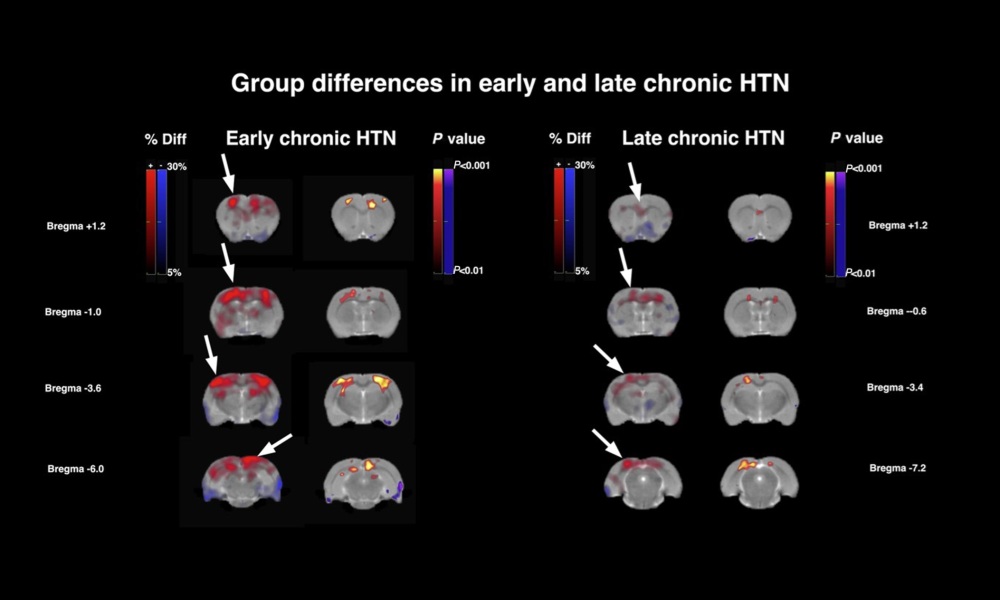
Figure 7. Brain regional glucose metabolism studied with 18F-fluorodeoxyglucose PET in chronic hypertension. Sections of group mean intensity–normalized 18F-fluorodeoxyglucose uptake are shown in the upper half of the figure, overlaid on an anatomical reference MRI. Color scale is in multiples of the global mean μ. In the lower half, percentage differences and statistically significant differences between early and late chronic hypertension and age-matched controls are shown. Warm colors indicate higher, blue colors lower metabolism in hypertensive rats. Note the marked increase in metabolism in early chronic HTN (hypertension) of up to 30% in cortical areas (arrows) including motor and cingulate cortex and in parts of the hippocampus. In late chronic HTN the spatial distribution of affected regions is largely similar, but the effect size is markedly reduced as compared with early chronic HTN.
Conclusion
- Chronic hypertension caused behavioral deficits associated with frontal cortex function.
- Results highlight stage-dependent responses to continuous microvascular stress and wounding by hypertension.
- Early chronic responses included a fast recruitment of activated microglia to the blood vessels, immigration of peripheral immune cells, blood–brain barrier breakdown and an energy-demanding hypermetabolic state. Vascular adaptation mechanisms were observed in later stages and included angiogenesis and upregulation of cellular adhesion molecules and extracellular matrix.
- Among the top upregulated genes in blood vessels, Igfbp-5 was identified, which attenuates protective insulin-like growth factor 1 signaling.
- This study provides new insights into the mechanisms underlying the pathobiology of hypertension and highlights its dependence on disease stage.
- This groundwork regarding the mechanisms will be helpful for basic and clinical research to identify stage-dependent markers in the human disease course, investigate stage-dependent interventions besides blood pressure lowering, and better understand the relationship between poor vascular health and neurodegenerative diseases.
Full article on ahajournals.com

How can we help you?
Don't hesitate to contact us for technical information or to find out more about our products and services.
Get in touch