The post-acute phase of SARS-CoV-2 infection in two macaques species is associated with signs of ongoing virus replication and pathology in pulmonary and extrapulmonary tissues
2020.11.05.
Kinga P. Böszörményi et al., BioRXiv, 2020
Summary
SARS-CoV-2 is a coronavirus that sparked the current COVID-19 pandemic. To stop it, effective and safe vaccines, and antiviral therapies are urgently required. To facilitate the preclinical evaluation of intervention approaches, relevant animal models need to be developed and validated. Rhesus macaques (Macaca mulatta) and cynomolgus macaques (Macaca fascicularis) are widely used in biomedical research and serve as models for SARS-CoV-2 infection, but this is the first controlled comparative study investigating which species of them is best suited to examine specific aspects of COVID-19. This study analysed replication and symptoms for three weeks after infection. Pulmonary lesions were detected on CT images acquired with MultiScan™ LFER PET/CT. Elevated body temperature and decreased in physical activity was also observed. Results show that both rhesus and cynomolgus macaques represent valid models for COVID-19 prophylactic and therapeutic treatments.
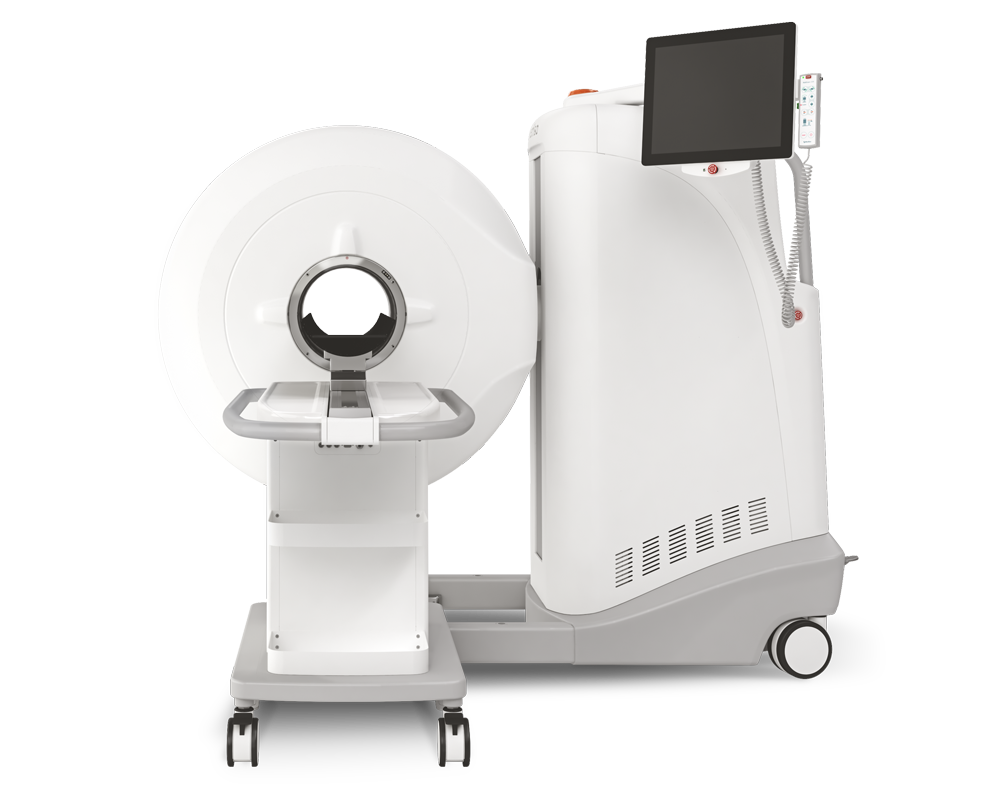
Results from MultiScan™ LFER PET/CT
CT imaging provides a valuable tool to specifically monitor the progression of COVID-19-related lung pathology during the entire course of the study. Respiratory-gated CT scans were performed on Day0, 2, 4, 6, 8, 10, 12, 14, 16, 22 post-infection to monitor lung pathology. A semi-quantitative scoring system for chest CT evaluation was used to estimate SARS-CoV-2-induced lung disease; maximum score of 35 could be reached per timepoint.
Scans revealed that:
- All macaques show levels of pneumonia
- Detected different types of lesions: A) ground glass opacities, B) consolidations, and C) crazy paving patterns (Figure 1)
- Around days 8 and 10 pi., lesions were manifest in all animals, and in several macaques the coverage had increased
- Cumulative CT scores increased and no difference was observed between rhesus and cynomolgus macaques

Further results showed:
- Both groups of animals, the body temperature was significantly higher during the first two weeks after infection
- Significantly lower activity in all four rhesus macaques during the first period after infection, while this difference in cynomolgus macaques was less obvious
- Antibody response became evident between day 10 and 12 pi., IgG level continued to rise for several days (development of IgM titers was barely detected)
- Certain cytokines increased in the plasma of both macaque species
How can we help you?
Don't hesitate to contact us for technical information or to find out more about our products and services.
Get in touch