Stereotactical normalization with multiple templates representative of normal and Parkinson‑typical reduction of striatal uptake improves the discriminative power of automatic semi‑quantitative analysis in dopamine transporter SPECT
2023.03.29.
Apostolova et al., EJNMMI Physics, 2023
Background
The specific binding ratio (SBR) of 123I-FP-CIT in the putamen is widely used to support the interpretation of dopamine transporter (DAT) SPECT. Automatic methods for computation of the putamen SBR often include stereotactical normalization of the individual DAT-SPECT image to an anatomical standard space. This study compared using a single 123I-FP-CIT template image as target for stereotactical normalization versus multiple templates representative of normal and different levels of Parkinson-typical reduction of striatal 123I-FP-CIT uptake.
Methods
1702 clinical 123I-FP-CIT SPECT images were stereotactically normalized (affine) to the anatomical space of the Montreal Neurological Institute (MNI) with SPM12 either using a single custom-made 123I-FP-CIT template representative of normal striatal uptake or using eight different templates representative of normal and different levels of Parkinson-typical reduction of striatal FP-CIT uptake with and without attenuation and scatter correction.

Fig. 1 Templates of the 123I-FP-CIT distribution volume ratio (DVR) in MNI space. The templates are representative of normal striatal signal (left column), moderate (middle columns) and strong (last column) Parkinson-typical reduction of striatal uptake with and without attenuation and scatter correction (ACSC). Each template was generated from twelve randomly selected DAT-SPECT images. The 123I-FP-CIT template representative of normal striatal signal with attenuation and scatter correction (upper left) was used for single template stereotactical normalization
In the latter case, SPM finds the linear combination of the multiple templates that best matches the patient’s image. The putamen SBR was obtained using hottest voxels analysis in large unilateral regions-of-interest predefined in MNI space. The histogram of the putamen SBR in the whole sample was fitted by the sum of two Gaussians. The power to differentiate between reduced and normal SBR was estimated by the effect size of the distance between the two Gaussians computed as the differences between their mean values scaled to their pooled standard deviation.
SPECT imaging
SPECT had been performed between December 2008 and January 2020 according to common procedures guidelines [19, 20] with four different cameras: Siemens e.cam dual head camera equipped with low-energy-high-resolution collimators, Siemens Symbia TruePoint dual head camera with low-energy-high-resolution collimators, Siemens Symbia TruePoint with fan-beam collimators, and Mediso AnyScan® TRIO triple head camera equipped with low-energy-high-resolution-high-sensitivity collimators in dual head mode. Detailed acquisition parameters are given in Table 1.

All SPECT images were reconstructed retrospectively using the iterative ordered-subsets-expectation–maximization algorithm with resolution recovery implemented in the HybridRecon-Neurology tool of the Hermes SMART workstation v1.6 with parameter settings recommended for FP-CIT SPECT by Hermes (5 iterations, 15/16 subsets for 120/128 views, postfiltering with 3-dimensional Gaussian kernel of 7 mm full-width-at-half-maximum, uniform attenuation correction with narrow-beam attenuation coefficient 0.146/cm, simulation-based scatter correction, resolution recovery with a Gaussian model).
Results
Among the 36 cases used for template generation, the putamen SBR (multiple templates, hottest voxels analysis, minimum of both hemispheres) in the 12 images with normal striatal signal was 1.896 ± 0.313, range 1.420–2.409, corresponding to the range ≥ 65th percentile in the whole data set, it was 0.663 ± 0.148, range 0.499–1.034, corresponding to 15th–45th percentile, in the 12 images with moderate reduction, and it was 0.282 ± 0.034, range 0.228–0.333, corresponding to ≤ 5th percentile, in the 12 images with strong reduction of the striatal signal. This suggests that the range of striatal 123I-FP-CIT uptake encountered in clinical practice was adequately covered by the multiple templates.
There was no major failure of stereotactical normalization according to visual inspection when using multiple templates as target. There were two major failures (0.1%) with the single template as target (Additional file 1: Fig. S1). These two DAT-SPECT were excluded from the further analyses. This resulted in the inclusion of 1702 DAT-SPECT from 1675 different patients (43.1% females). The age at the time of DAT-SPECT was 66.4 ± 11.5 years (range 20–90 years). The activity dose of 123I-FP-CIT injected for these DAT-SPECT was 194 ± 21 MBq (range 115–291 MBq).
The general linear model for repeated measures revealed a significant between-subjects effect of the camera on the putamen SBR (F = 16.4, p < 0.0005). The mean putamen SBR was highest in scans acquired with the AnyScan® TRIO with LEHRHS collimators and lowest in scans acquired with the E.Cam with LEHR collimators. On average, putamen SBR were 0.254 larger in the images acquired with the AnyScan Trio with LEHRHS collimators than in the images (from different patients) acquired with the e.cam with LEHR collimators.
Full article on EJNMMI Physics
-
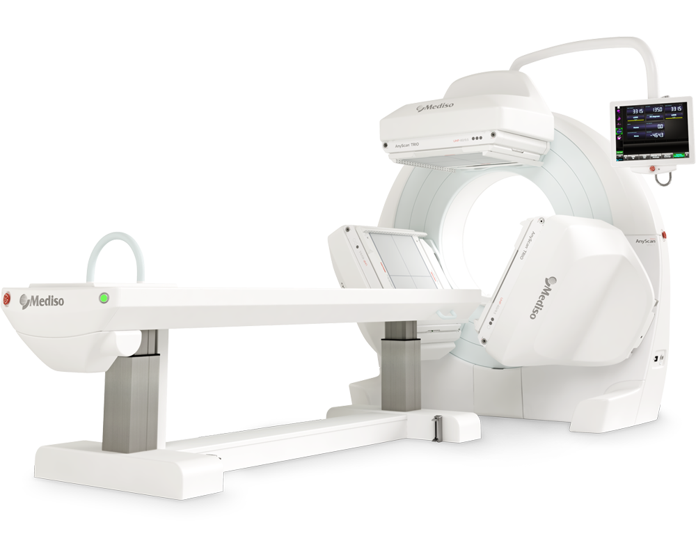
AnyScan® TRIO SPECT
Ultra-fast Triple-NaI-Detector SPECT for wide range of clinical applications
-
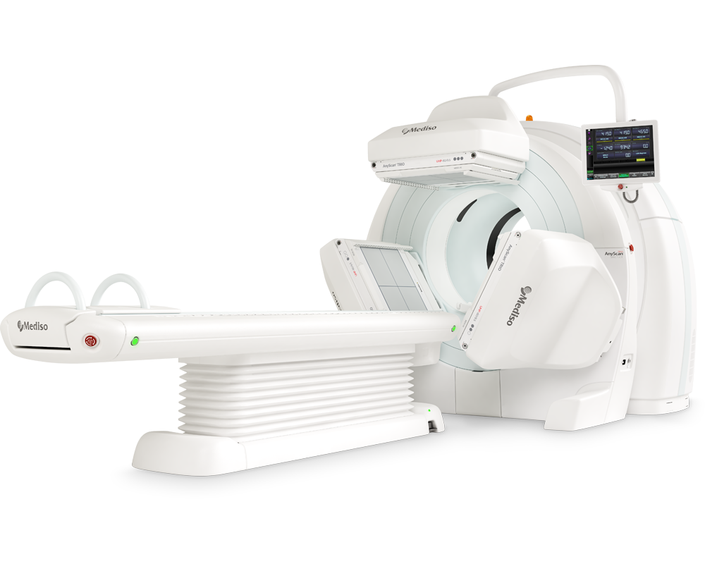
AnyScan® TRIO SPECT/CT
Ultra-fast Triple-NaI-Detector SPECT system for wide range of clinical applicati...
-
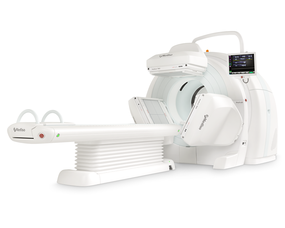
AnyScan® TRIO SPECT/CT/PET
An integrated triple-detector SPECT/CT and PET/CT system in a single-room instal...
-
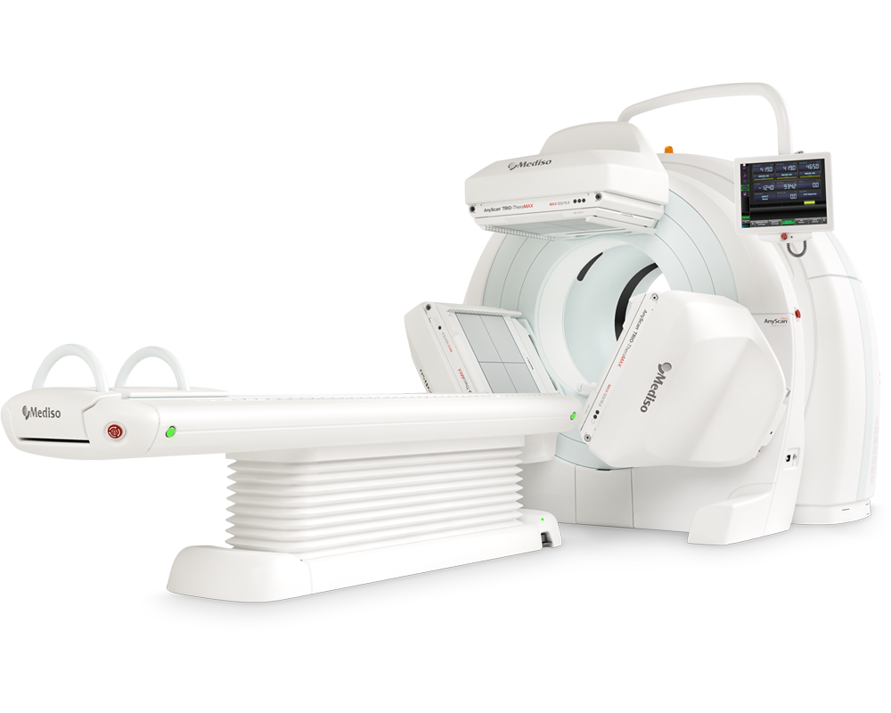
AnyScan® TRIO-TheraMAX SPECT/CT
Theranostic and Diagnostic Imaging with MAXimum Performance


How can we help you?
Don't hesitate to contact us for technical information or to find out more about our products and services.
Get in touch