Responses to acute infection with SARS-CoV-2 in the lungs of rhesus macaques, baboons and marmosets
2020.12.18.
Dhiraj Kumar Singh, Bindu Singh, Deepak Kausha;
Nature Microbiology, 2021
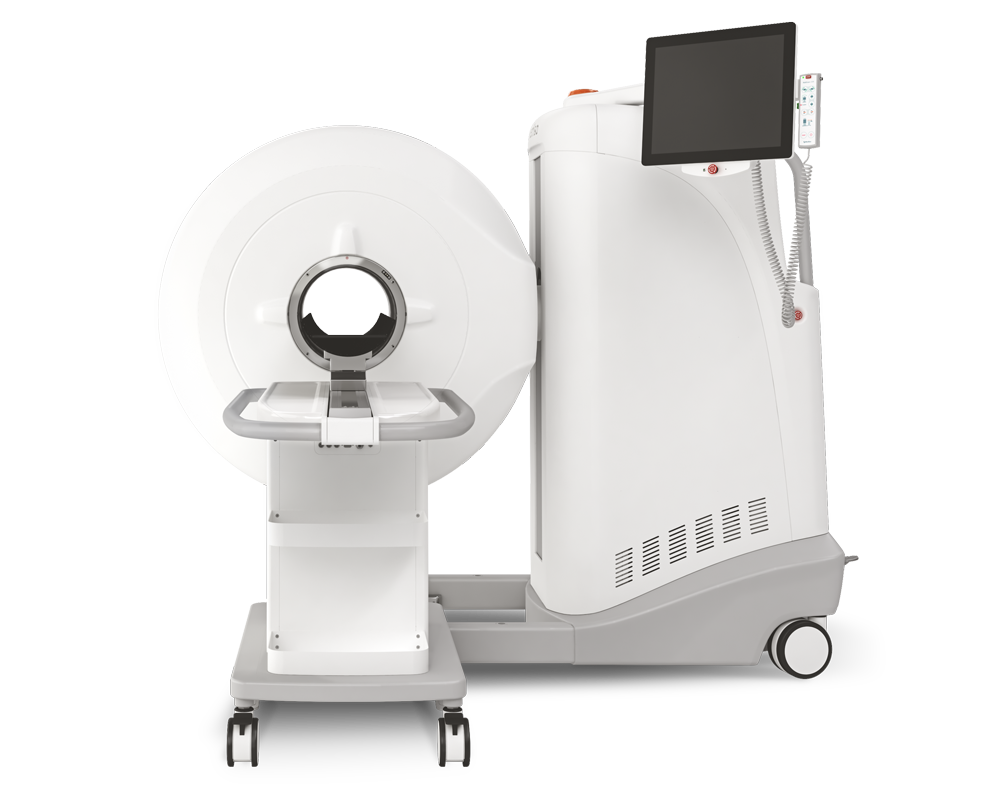
Southwest National Primate Research Center in Texas Biomedical Research, San Antonio has purchased Mediso’s MultiScan® LFER PET/CT scanner in early 2020. Mediso USA proudly post that Professor Deepak Kaushal and his co-workers used MultiScan® LFER PET/CT in their recently reported comprehensive work about the course of SARS-CoV-2 infection in nonhuman primate models.
MultiScan® LFER PET/CT features a 20 cm axial and 15 cm transaxial field of view with a 26 cm bore opening which allows the researcher to scan non-human primates (NHP) with sub-mm PET and even 150 um CT resolution.
Summary
The authors compare SARS-CoV-2 infection in three species of experimentally infected NHPs (rhesus macaques, baboons, and marmosets). They used a wide variety of methods to describe the course of disease such as clinical parameters of viral infection, viral RNA and viral protein detection, immune response, X-ray, CXR scoring, CT scanning and pathology.
Their results show all NHPs can be infected with SARS-CoV-2 but exhibit differential progression to COVID-19. Baboons exhibit moderate to severe pathology, macaques exhibit moderate pathology and marmosets exhibit mild pathology. They also summarize that rhesus macaques and baboons develop different, quantifiable disease attributes making them immediately available essential models to test new vaccines and therapies.
Results from MultiScan® LFER PET/CT
The report shows the importance of state-of-the-art, non-invasive imaging – cone beam CT scanning, and the application of innovative algorithms to identify the extent of lung involvement in pneumonia in developing models of COVID-19. CT image analysis provided a quantifiable metric data which enables testing efficacy of vaccines or the impact of therapeutic intervention. Lung hyperdensity and the CT abnormality score was used as a metric parameter to follow the onset of the disease.
Each NHP species was infected and followed over a 3-day period to describe the early signs of infection. Cone-beam CT scans showed evidence of moderate pneumonia, which progressed over 3 days (Figure 1). CT images were analyzed using the segmentation tool in VivoQuant (Invicro, Boston,MA). Lung hyperdensity and the CT abnormality score were used as a metric parameter to follow the course of the disease.

Figure 1. a) 3D Reconstruction of ROI volume representing the location of the lesion. b-d) represent images for quantification of the lung lesion with the green area representing normal intensity lung voxels (-850 HU to -500 HU), while red areas represent hyperdense voxels (-490 HU to 500 HU). (Image courtesy of Dr. Deepak Kaushal, Texas Biomedical Research)
They also performed detailed imaging of macaques in a 12-day longitudinal study. Similar to the acute study. imaging revealed a significant, progressive increase in the volume of lung involved in pneumonia at 6 dpi, which normalized by 12 dpi.
These results suggest that pneumonia in some older macaques may persist longer than in younger animals. Although there were several smaller changes observed in older animals, old and young animals both resolved the infection.
How can we help you?
Don't hesitate to contact us for technical information or to find out more about our products and services.
Get in touch