Quantitation of the A2A Adenosine Receptor Density in the Striatum of Mice and Pigs with [18F]FLUDA by Positron Emission Tomography
2022.04.22.
Daniel Gündel et al, Pharmaceuticals, 2022
Summary
The cerebral expression of the A2A adenosine receptor (A2AAR) is altered in neurodegenerative diseases such as Parkinson’s (PD) and Huntington’s (HD) diseases, making these receptors an attractive diagnostic and therapeutic target. The aim of this study was to further investigate the pharmacokinetic properties in the brain of the recently developed A2AAR–specific antagonist radiotracer [18F]FLUDA. For this purpose, dynamic PET studies of healthy mice and rotenone–treated mice (rotenone-induced PD group) were analysed retrospectively, and dynamic PET studies with healthy pigs were also conducted. Mouse brain time–activity curves were analysed to calculate the mean residence time (MRT) by non–compartmental analysis. Binding potential (BPND) of [18F]FLUDA was evaluated using the simplified reference tissue model (SRTM). For the pig studies, a Logan graphical analysis was performed to calculate the radiotracer distribution volume (VT) at baseline and under blocking conditions with tozadenant (A2AAR antagonist). The MRT of [18F]FLUDA in the striatum of mice was decreased by 30% after treatment with the istradefylline (another A2AAR antagonist). Mouse results showed the highest BPND (3.9 to 5.9) in the striatum. SRTM analysis showed a 20% lower A2AAR availability in the rotenone–treated mice compared to the control–aged group. Tozadenant treatment significantly decreased the VT (14.6 vs. 8.5 mL / g) and BPND values (1.3 vs. 0.3) in pig striatum. This study confirms the target specificity and a high BPND of [18F]FLUDA in the striatum. In conclusion, [18F]FLUDA is a suitable tool for the non–invasive quantitation of altered A2AAR expression in neurodegenerative diseases such as PD and HD, by PET.
Results from nanoScan® PET/MRI
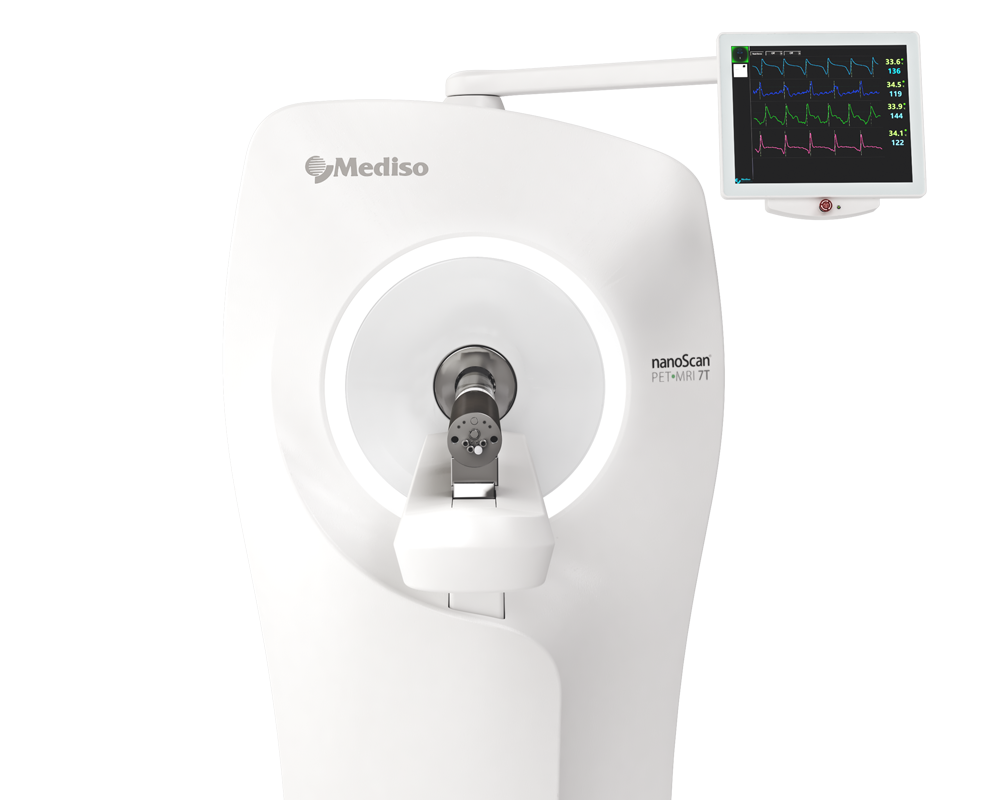
Wild–type C57BL/6JRj mice were divided into two groups and treated five days a week for two months either with vehicle (control group) or with rotenon (treatment group; 5 mg/kg daily dose). The CD–1 mice were divided into three groups: a baseline group with intravenous vehicle injection and a pre–treatment group with a blocking agent administered by intravenous injection (istradefylline, 1.0 mg/kg or tozadenant, 2.5 mg/kg) eight or fifteen minutes prior to radiotracer injection. The animals received an injection of [18F]FLUDA into a tail vein (3.1–9.7 MBq in 150 µL, for CD–1 mice; 3.7–8.2 MBq in 150 µL for C57BL/6JRj mice). A 60 min PET/MR scan was initiated (Mediso nanoScan®, Budapest, Hungary) at the time of tracer injection. Subsequently, a T1–weighted gradient–echo sequence (GRE, repetition time = 20 ms, echo time = 6.4 ms) was performed for whole body attenuation correction and anatomical orientation. Additionally, PET data were corrected for random coincidences, dead time, and scatter. The list mode data were sorted into sinograms using a framing scheme of 12 x 10 s, 6 x 30 s, 5 x 60 s, 10 x 300 s. The reconstruction parameters were the following: 3D–ordered subset expectation maximization (OSEM), four iterations, six subsets, energy window = 400–600 keV, coincidence mode = 1–5.
Brain volume of interest (VOI) analysis for mouse experiments were performed with PMOD software. Kinetic analysis revealed that:
- treatment with istradefylline, but not tozadenant, significantly reduced the area under the curve (AUC) in the murine striatum
- pre–treatment with tozadenant was without effect on the kinetic parameters calculated for the target region striatum or for the reference region cerebellum

- in contrast, the pre–treatment with istradefylline tended to shorten the time–to–peak and to diminish the TAC peak value in the striatum to a level comparable with the values in the cerebellum. Hence, the mean residence time (MRT) was significantly reduced in the striatum. pre–administration of istradefylline did not alter the MRTs in the cerebellum, validating its use as a reference region.

- mean parametric BPND maps showed high total binding of [18F]FLUDA in the mouse striatum and a complete blocking by pre–treatment with istradefylline; the striatal BPND declined from 3.9 ± 1.2 to zero.

- mean parametric BPND maps suggested a 20% lower striatal BPND in the rotenone–treated mice compared to the control group

Full article on www.mdpi.com
How can we help you?
Don't hesitate to contact us for technical information or to find out more about our products and services.
Get in touch