New workflows and algorithms of bone scintigraphy based on SPECT-CT
2012.10.28.
Peter Bandi et al., IEEE, 2012
Abstract
Gold standard bone scintigraphy workflow contains acquisition of planar anterior and posterior images and if necessary, additional SPECTs as well. Planar acquisitions are time consuming and not enough for accurately locating hotspots. Current paper proposes a novel workflow for fast whole body bone SPECT scintigraphy. We present a novel stitching method to generate a whole body SPECT based on the SPECT projections. Our stitching method is performed on the projection series not on the reconstructed SPECTs, thus stitching artifacts are greatly reduced. Our workflow does not require any anterior-posterior image pairs, since these images are derived from the reconstructed whole body SPECT automatically. Our stitching method has been validated on real clinical data performed by medical physicians. Results show that our method is very effective for whole body SPECT generations leaving no signs of artifacts. Our workflow required overall 16 minutes to acquire a whole body SPECT which is comparable to the 60 minutes acquisition time required for gold standard techniques including planar images and additional SPECT acquisitions.
Patient images
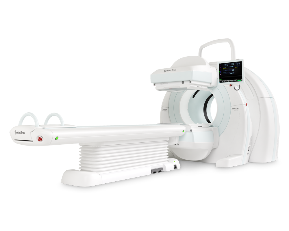
We have collected 36 anonymized DICOM studies acquired by Mediso AnyScan® SC SPECT/CT multi-modal camera system. The studies contained the SPECT-CT acquisitions, the reconstructed CT and the SPECT projections. The SPECT images were acquired after injecting Tc-99m MDP radiopharmacon. The type of the collimator was LEGP. The resolution of the SPECT projections was 256 [1] 256 pixels. One projection took 5 seconds to acquire. Overall 64 projections were acquired for one FOV and generally 2-6 FOVs were collected for a patient. The detector shape was octagonal having cut corners of a rectangular shape. The overlapping size of the detector views was 40 mm. All overlapping projections were spatially aligned and this alignment information was derived from the DICOM images, hence no registration was necessary. The average whole body SPECT acquisition took 16 minutes in case of 5 FOVs (5 ∗ 32 = 160 seconds / FOV). The CT values were Hounsfield corrected.
Methods
1) Stitching: We assumed that the input was a P set of partially overlapping S p projective SPECT image series of a patient. Each of these series consisted of 2D projective images acquired from different α angles: S p = {S pα|α ∈ R}, where R is the acquisition angle set. The dimensions of the projective images and the acquisition angle set were the same for all S p series. Spatial and rotational alignment of these images from different series was performed automatically based on DICOM tags
2) Artificial planar A-P image generation: We reconstructed the stitched whole body SPECT projections by applying the OSEMRRAC reconstruction method [7], [8]. The desired reconstructed SPECT axial matrix size was 128 × 128 with 0.4mm pixel spacing. The reconstructed CT was resampled and cropped to have the same size as the above SPECT matrix size. The interpolation method for the CT resampling was tri-cubic. This step increased the speed of further CT processing and allowed us to directly calculate attenuation coefficients on the SPECT voxel level. Multi-threshold was performed on the resampled CT to generate a material map for the attenuation correction.
The reconstructed whole body SPECT was projected to anterior and posterior direction to generate the artificial planar pairs. During the projection the above CT based material map was used as well. For results of an artificial planar anterior and posterior image pair projected from the WB SPECT see Fig. 3.

Fig. 3. Artificial planar anterior (left) and posterior (right) image pairs generated from a whole body stitched and reconstructed SPECT. Note that no stitching regions can be discovered on the image pairs.
Validation
Validation was based on visual inspection and performed by two medical physicians in InterView™ FUSION clinical evaluation software developed by Mediso. The validation process covered two main questions: quality of stitching and quality of the artificial planar images.

Fig. 4. Image stitching techniques performed before reconstruction. Left: average, Middle: cross-fade, Right: our method. Note the stitching effect at overlapping regions on average and cross-fade stitched images.
Results
The planar image projections did not include noticeable stitching artifacts based on our method and they appeared to be a useful aid for fast localization during the evaluation. For an example layout of the evaluation see Fig. 5.

Fig. 5. Example layout for evaluating our whole body SPECT studies. First two images show the artificial anterior - posterior image pairs. Middle three images show the axial-sagittal-coronal views of the fused whole body SPECT-CT. Last image shows the volume rendering view of the fused whole body SPECT-CT. Note the synchronized cursor position representing a hotspot in the shoulder.
Conclusions
We have proposed a new SPECT-CT acquisition workflow with a novel stitching algorithm to provide whole body SPECT images for bone scans. The difference comparing to the gold standard workflows appears on two levels: On one hand we do not acquire planar scans at all, but in return we acquire SPECT scans that are stitched to a whole body SPECT before reconstruction. Our stitching method is very accurate and fully automated due to the approach of stitching before reconstruction on the level of projections. We derive artificial planar images from the reconstructed SPECT to aid localization during evaluation. On the other hand the overall time to acquire a SPECT scan series covering the whole body takes average 16 minutes which is comparable to present gold standard workflows including a planar scan pair and an additional SPECT in 45-60 minutes. This approach can significantly decrease the time to per- form a bone scan in the daily routine which helps maximizing the number of scans per day.
Full article on IEEE Xplore.
How can we help you?
Don't hesitate to contact us for technical information or to find out more about our products and services.
Get in touch