Renal PET-imaging with 11C-metformin in a transgenic mouse model for chronic kidney disease
2016.06.23.
Lea Pedersen et al, EJNMMI Research, 2016
Summary
Background: Organic cation transporters (OCTs) in the renal proximal tubule are important for the excretion of both exo- and endogenous compounds, and chronic kidney disease (CKD) alter the expression of OCT. CKD can be studied in a mouse model with TGFβ-induced chronic kidney disease (RenTGF-β1). These mice express proximal tubular dysfunction, tubular basement membrane thickening, interstitial fibrosis, and proteinuria when they are approximately 4-month old. Metformin is a well-known substrate for OCT, and recently, it was demonstrated that positron emission tomography (PET) with 11C-labelled metformin is a promising approach to evaluate the function of OCT. The aim of this study is therefore to examine renal pharmacokinetics of 11C-metformin and expression of OCTs in the RenTGF-β1 mouse model.
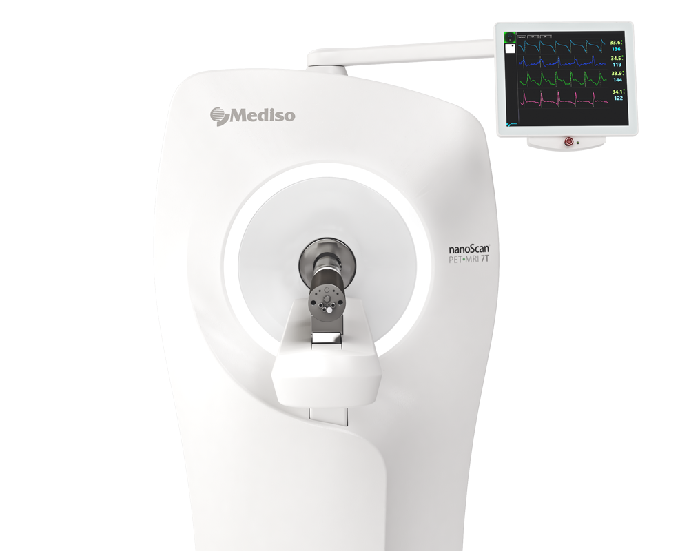
Methods: Age- and sex-matched RenTGF-β1 (Tg) and wildtype (WT) mice received an iv bolus of 11C-metformin followed by 90-min dynamic PET and MRI scan using nanoScan PET/MRI. PET data were analysed using a one-tissue compartment model. As in the proximal tubule of mice, OCT2 (and to a lesser degree OCT1) mediates basolateral uptake of metformin, whereas the multidrug and toxin extrusion protein 1 (MATE1) is responsible for excretion into the tubule lumen in a H+-coupled electroneutral manner, renal protein abundance of OCT2 (by Western blot) as well as OCT1, OCT2, and MATE1 messenger RNA (mRNA) (by RT-PCR) was also examined.
Results: Protein expression of OCT2 was 1.5-fold lower in Tg mice compared to WT mice while OCT1 and MATE1 mRNA expression did not differ between the two groups. The influx rate constant of 11C-metformin in renal cortex (K1) was 2.2-fold lower in transgenic mice whereas the backflux rate constant (k2) was similar in the two groups, consistent with protein expression. Total body clearance (TBC) correlated within each group linearly with K1.
In conclusion, this study demonstrates that both renal OCT2 expression and 11C-metformin uptake are reduced in CKD mice. This potentially makes 11C-metformin valuable as a PET probe to evaluate kidney function.
Results from nanoScan® PET/MRI
For the kinetic modelling study RenTGF-β1 transgenic (Tg) mouse strain was used: mutated porcine TGF-β1 is expressed under the control of the Ren-1c promoter. To further investigate the relative importance of OCT1/2 and MATE1, total body clearance (TBC) was calculated. This included controls at baseline and after treatment with cimetidine (an OCT1/2 inhibitor) or pyrimethamine (a MATE1 inhibitor) and OCT1/2 knock-out mice.
A single bolus of of 11C-metformin (7.7 ± 4.0 MBq/mouse) was injected via the catheter to Tg females (n = 8; 4-5 month old) and age- and sex-matched wildtype (WT) littermates (n = 5) and 90min dynamic PET scan was started with nanoScan PET/MRI. PET acquisition was followed by anatimical MRI scan. Body temperature was kept at 36–37 °C throughout the intervention, and respiration frequency was monitored.
Multiple regions of interest (ROIs) were placed on the renal cortex, heart, and liver using PMOD version 3.5 creating a volume of interest (VOI). Half-moon shaped renal cortex VOIs were defined on an average image of the 33 PET frames. Subsequently, it was verified that the VOIs were placed within the renal cortex in each frame. Hepatic VOIs were drawn on the first 25 frames where it can easily be identified and averaged. The heart was used as an image-derived input function. Frames from the first 20 s were averaged and ROI circles with a diameter of 15 pixels were placed on the six most intensive slices in the middle of the heart. Correct positioning of the liver and heart VOIs were checked in each time frame and adjusted if needed. Time-activity curves (TACs) were generated from the VOIs.
Dynamic PET data for each kidney cortex were fitted using a one-tissue compartment model with two parameters: K1, the influx rate constant (from blood into the tissue compartment) and k2, the backflux rate constant (out of the tissue compartment). The kinetic parameters were estimated by minimizing the residual sum of squares (RSS) using the Levenberg-Marquard algorithm. A single mouse was excluded from the analyses because the model fit yielded non-physiological parameter estimates.
TBC during 90 min was calculated based on the image-derived input function from the following equation:

where ID is injected dose relative to kilogram body weight and AUC(0–90 min) represents Area Under the Curve of the image-derived input function up to 90 min.
Results show:
- The renal distribution of 11C-metformin peaked from 1–5 min and decreased towards 90 min due to extensive urinary excretion

Coronal whole body PET with 11C-metformin merged with T1-weighted MRI-sequence in a WT mouse (upper panel) and a Tg mouse (lower panel). The projection is posterior to the liver and heart. Radioactivity in the kidneys peaks from 1 to 5 min and decreases towards 90 min because of extensive urinary excretion. Scale bar to the left represents standard uptake value (SUV) 0–5

Time-activity curves of 11C-metformin in the kidneys of Tg and WT mice
- TBC of 11C-metformin was determined using the image-derived input function and was 1.8-fold lower in Tg compared to WT mice. No difference was observed between the left and right kidney; consequently, rate constants were expressed as means of both kidneys.
- The influx rate constant in renal cortex (K1) was 2.2-fold lower in Tg mice. k2 did not differ significantly between the two groups (data not shown).

Kinetic analysis of dynamic PET data. A: TBC of 11C-metformin in WT (n = 5) and Tg (n = 8) mice determined from image-derived input function. B: K1 in renal cortex determined from compartmental analysis
- Ablation of OCT1/2 or pre-treatment with cimetidine had a profound lowering effect on TBC whereas inhibition of MATE1 by pyrimethamine slightly increased TBC albeit insignificantly

TBC of 11C-metformin in RenTGF-β1 mice (Tg), OCT1/2 KO mice, or wildtype mice treated with an OCT1/2 inhibitor (cimetidine) or MATE1 inhibitor (pyrimethamine), respectively, relative to the appropriate control mice
Full article on ejnmmires.springeropen.com
Wie können wir Ihnen behilflich sein?
Bitte kontaktieren Sie uns für technische Informationen und Unterstützung jeglicher Art in Zusammenhang mit unseren Entwicklungen und Produkten.
Kontaktformular