Effects of isoflurane anaesthesia depth and duration on renal function measured with [99mTc]Tc-mercaptoacetyltriglycine SPECT in mice
2024.01.05.
Fabian Schmitz-Peiffer et al., EJNMMI Res., 2024
Summary
In a study involving 86 severe combined immunodeficient (SCID) mice, researchers investigated the impact of different levels of isoflurane anesthesia and handling procedures on kidney scintigraphy using dynamic single photon emission computed tomography (SPECT). They adjusted isoflurane concentrations for low and deep anesthesia based on respiratory rate and compared single bed versus 3-mouse bed hotel setups. Results showed that deep anesthesia and prolonged low anesthesia led to delays in blood clearance and renal influx, as well as later time-to-peak values. Conversely, low anesthesia with high respiratory rates and short durations yielded more representative data with lower variance. These findings suggest that careful control of anesthesia depth and duration is crucial for obtaining accurate renal scintigraphy results in preclinical studies.
- Preclinical evaluation of medicinal products, particularly diagnostic and therapeutic tracers, is pivotal in their development and eventual approval. In nuclear medicine, optimizing peptide-based tumor therapies while minimizing kidney uptake and associated toxicities poses a significant challenge. Therefore, establishing reliable preclinical tests for assessing kidney toxicity is crucial for advancing new therapeutic radiopharmaceuticals.
- Renal scintigraphy, a non-invasive imaging technique, plays a central role in measuring parameters such as glomerular filtration rate (GFR) and renal blood flow (RBF). Utilizing 99mTechnetium mercaptoacetyltriglycine ([99mTc]Tc-MAG3) as a tubular tracer, renal scintigraphy provides robust quantification of renal function. However, various physiological factors and external influences, including anesthesia, can affect its efficacy.
- Isoflurane, a widely used inhalation anesthetic in preclinical research, offers advantages such as rapid onset and controllability. Nonetheless, its impact on the pharmacokinetics of radiotracers like [99mTc]Tc-MAG3 remains under investigation. Understanding this interaction is critical for accurately assessing renal function impairment in drug development.
- Thus, this study aimed to elucidate the effects of different depths of isoflurane anesthesia on renal excretion parameters using dynamic single photon emission computed tomography (SPECT) imaging in mice. Additionally, we aimed to investigate the influence of anesthesia duration by comparing single bed versus multiple mouse bed setups, recognizing the importance of both depth and duration of anesthesia in renal function assessments.
Methods
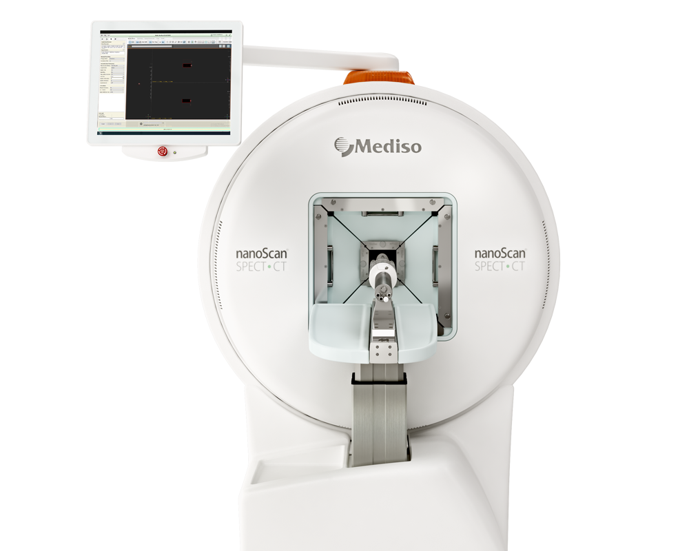
SCID mice, vital for tumor modeling in oncology, were utilized exclusively in this study. Animals were housed under controlled conditions following a 12-hour light-dark cycle. Renal scintigraphy was conducted using NanoSPECT/CTplus (Mediso, Hungary/Bioscan, France) equipment, equipped with high-resolution mouse apertures for precise imaging. The study design for assessing the impact of anesthesia depth and duration in mice is depicted in Figure 1.

Fig. 1 Investigation of isoflurane anaesthesia depth controlled by respiratory rates (low = 80–90/min, deep = 40–45/min) in a single mouse bed. [99mTc]Tc-MAG3 injection started approx. 10 min after anaesthesia induction. b Investigation of isoflurane anaesthesia duration at low anaesthesia in comparison between mice imaged in a single mouse bed and in a 3-mouse bed hotel. At the time of tracer injection, anaesthesia duration was approx. 30 min for mice in hotel position 1, 20 min in position 2, and 10 min in position 3 and single mouse bed. Mice created with Biorender.com
Investigation of Isoflurane Anesthesia Depth:
- Seven male SCID mice aged 5 months were studied.
- Imaging was performed under low and high isoflurane concentrations, one week apart.
- Isoflurane concentrations ranged from 1-2%, adjusted based on respiratory rates.
- Respiratory rates of 80-90/min and 40-45/min were used for low and deep anesthesia, respectively.
- Electrocardiography (ECG) monitoring was conducted using neonatal electrodes.
- To avoid carry-over effects, half of the mice were first examined under low isoflurane concentration, while the other half started with high concentration.
- Examinations were performed at the same time of day to eliminate potential circadian rhythm effects.
Investigation of Isoflurane Anesthesia Duration:
- Twelve female SCID mice aged 3 months were studied.
- Imaging was conducted in a heated single mouse bed.
- Respiratory rate was maintained at 80-90 breaths/min.
- Tracer injection followed SPECT start approximately 10 min after anesthesia induction.
- Sixty-seven female SCID mice aged 3-4 months were studied.
- Imaging was performed in a heated 3-mouse bed hotel.
- Isoflurane concentration was uniformly distributed among bed positions.
Tracer injection was staggered among mice based on catheter placement, with varying durations of anesthesia (30 min for position 1, 20 min for position 2, and 10 min for position 3) at the time of injection.
Results from nanoScan® SPECT/CT
Renal scintigraphy by semi-stationary SPECT/CT
During renal scintigraphy, mice were anesthetized with isoflurane for catheterization, tracer injection, and SPECT/CT acquisition. A 30 G cannula and a 0.28 × 0.61 mm catheter filled with 2 I.U. heparin per ml 0.9% NaCl were used for intravenous tail injection of approximately 28 MBq [99mTc]Tc-MAG3. SPECT acquisition, following a low-dose CT scan, consisted of 68 reconstructed images over 35 minutes, with each frame lasting 20 or 50 seconds. In the 3-mouse bed hotel setup, where mice were injected consecutively, dynamic data acquisition began with 78 reconstructed images over 38 minutes to ensure detection of Tmax for all mice. Additional anesthesia time varied, with 10 minutes for mice in single beds and position 3 of the hotel, approximately 30 minutes for mice initially placed in position 1 of the hotel, and around 20 minutes for position 2. After completion of the procedure, tail catheters were removed, and mice were allowed to wake up in a heating box before returning to their housing group.
Quantification and statistical analysis
The SPECT device undergoes regular calibration by the manufacturer to ensure accuracy in high voltage, energy, count rate linearity, energy resolution, and spatial uniformity. Cross-calibration with a dose calibrator is conducted for each nuclide, generating absolute values in kBq with sufficient quantitative accuracy. Aortic and renal uptake kinetics of [99mTc]Tc-MAG3 were assessed by windowing the data and defining volume-of-interest (VOI) contours for each kidney, encompassing renal cortex, medulla, and the pelvicalyceal system. Time-to-peak (Tmax), T50 (50% clearance), T25 (75% clearance), and blood excretion half-life (aorta 50% clearance) were calculated. Data are presented as median, interquartile range, minimum, and maximum, depicted as boxplots. Statistical comparisons were made using the Wilcoxon test for paired groups (e.g., low vs. deep anesthesia) and the Mann–Whitney U test for unpaired groups (e.g., single bed vs. hotel). Significance was assumed at p < 0.05. Repeated blood sampling for serum creatinine determination was not feasible due to the low total blood volume of mice in this longitudinal study.

Fig. 2 Maximum intensity projection of a semi-stationary dynamic SPECT acquisition fused with whole-body CT of mice acquired in a 3-mouse bed hotel with 22 mm scan range (high resolution rat apertures) after intravenous injection of [99mTc]Tc-MAG3. The color bar represents kBq. b SPECT acquisition in a single mouse bed with 14 mm scan range (high resolution mouse apertures) and manual contouring of a volume-of-interest (VOI) to quantify [99mTc]Tc-MAG3 uptake
Due to the anatomical positioning of the right kidney being more cranial than the left in mice, effectively capturing both kidneys within the limited scan range of 14 mm using mouse apertures can be challenging. Therefore, rat apertures with a larger transaxial field of view (22 mm) are more suitable for renal scintigraphy using semi-stationary SPECT in mice. Figure 2 illustrates the utilization of both rat and mouse apertures, demonstrating comparable image resolution between the two.
Effects of isoflurane anaesthesia depth on renal function
The depth of anesthesia, regulated based on respiratory rate rather than isoflurane concentration, significantly influenced renal function parameters during scintigraphy (Table 1). Low anesthesia with high respiratory rates led to shorter blood clearance half-life (T50aorta, p = 0.091) and significantly increased relative renal tracer influx rate (p = 0.018). There was a tendency for earlier time-to-peak (Tmax) under low anesthesia (p = 0.063), while T50 and T25 did not significantly differ. Heart rate showed no significant impact on renal tracer kinetics. However, data variance increased during deep anesthesia.
Different parameters during renal [99mTc]Tc-MAG3 scintigraphy in single mouse bed examinations as influenced by the depth of anaesthesia controlled by respiratory rate

Aorta tracer excretion half-life is expressed as T50aorta in seconds and kidney uptake as time-to-peak (Tmax), T50 (50% clearance) and T25 (75% clearance) in minutes. Each set of data includes the median, interquartile range [IQR], min–max and number of animals

Fig. 3 Time-to-peak (Tmax) of [99mTc]Tc-MAG3 kidney uptake during low and deep anaesthesia in single mouse bed examinations (p = 0.063)
Effects of single or multiple mouse beds on renal function
The positioning of mice in single or multiple beds within the hotel setup significantly impacted renal function parameters during scintigraphy. Mice in position 1, under anesthesia for the longest duration (approximately 30 minutes), exhibited a notably delayed time-to-peak (Tmax), T50, and T25 compared to mice in positions 2 and 3, as well as those in single beds (Table 2). Conversely, no significant differences were observed between single bed examinations and positions 2 or 3 within the hotel setup. However, within the hotel, significant differences were noted between position 1 and position 2 (with delayed T50 and T25; p < 0.01 each) and between position 1 and position 3 (with delayed Tmax, T50, and T25; p < 0.05 each). This disparity is visually represented in Figure 4, where the mouse in position 1 exhibited a distinct delay in Tmax compared to the other mice in the same acquisition.
[99mTc]Tc-MAG3 kidney kinetics compared between single mouse bed and 3-mouse bed hotel examinations

Aorta tracer excretion half-life is expressed as T50aorta in seconds and kidney uptake as time-to-peak (Tmax), T50 (50% clearance) and T25 (75% clearance) in minutes. Each set of data includes the median, interquartile range [IQR], min–max and number of animals
- #p < 0.05 between single versus position 1
- +p < 0.01 between position 1 versus 2
- ♦p < 0.05 between position 1 versus 3

Fig. 4 Example of [99mTc]Tc-MAG3 time activity curves of renal function (left and right kidney pooled, respectively) from three mice imaged simultaneously within a 3-mouse bed hotel after intravenous tracer injection. At time of tracer injection, duration of anaesthesia was approx. 30 min for position 1, 20 min for position 2, and 10 min for position 3. The mouse in position 1 showed delayed time-to-peak (Tmax), T50 (50% clearance) and T25 (75% clearance). Mice created with Biorender.com
In a conclusion, deep anesthesia in mice, characterized by reduced respiratory rates, led to adverse effects on renal function parameters, including prolonged blood clearance half-life, delayed renal tracer influx rate, and later Tmax, accompanied by increased variability in kinetics. Therefore, maintaining consistent respiratory rates and opting for low-depth anesthesia with high respiratory rates (80–90 rpm) during renal scintigraphy is recommended to ensure reliable data with minimal kinetic variability. Furthermore, prolonged anesthesia duration, particularly experienced by the first mouse in a 3-bed hotel setup, negatively impacted renal parameters. To mitigate this issue, investigations in multiple mouse bed hotels should ideally involve a maximum of two simultaneously examined mice, as individual adjustment to low anesthesia is challenging.
Full article on ncbi.nlm.nih.gov
Wie können wir Ihnen behilflich sein?
Bitte kontaktieren Sie uns für technische Informationen und Unterstützung jeglicher Art in Zusammenhang mit unseren Entwicklungen und Produkten.
Kontaktformular