Distributable, Metabolic PET Reporting of Tuberculosis
2023.04.03.
R M Naseer Khan et. al
this is a preprint at bioRxiv, 2023
ABSTRACT
Tuberculosis remains a large global disease burden for which treatment regimens are protracted and monitoring of disease activity difficult. Existing detection methods rely almost exclusively on bacterial culture from sputum which limits sampling to organisms on the pulmonary surface. Advances in monitoring tuberculous lesions have utilized the common glucoside [18F]FDG, yet lack specificity to the causative pathogen Mycobacterium tuberculosis (Mtb) and so do not directly correlate with pathogen viability. Here we show that a close mimic that is also positron-emitting of the non-mammalian Mtb disaccharide trehalose – 2-[18F]fluoro-2-deoxytrehalose ([18F]FDT) – can act as a mechanism-based enzyme reporter in vivo. Use of [18F]FDT in the imaging of Mtb in diverse models of disease, including non-human primates, successfully co-opts Mtb-specific processing of trehalose to allow the specific imaging of TB-associated lesions and to monitor the effects of treatment. A pyrogen-free, direct enzyme-catalyzed process for its radiochemical synthesis allows the ready production of [18F]FDT from the most globally-abundant organic 18F-containing molecule, [18F]FDG. The full, pre-clinical validation of both production method and [18F]FDT now creates a new, bacterium-specific, clinical diagnostic candidate. We anticipate that this distributable technology to generate clinical-grade [18F]FDT directly from the widely-available clinical reagent [18F]FDG, without need for either bespoke radioisotope generation or specialist chemical methods and/or facilities, could now usher in global, democratized access to a TB-specific PET tracer.
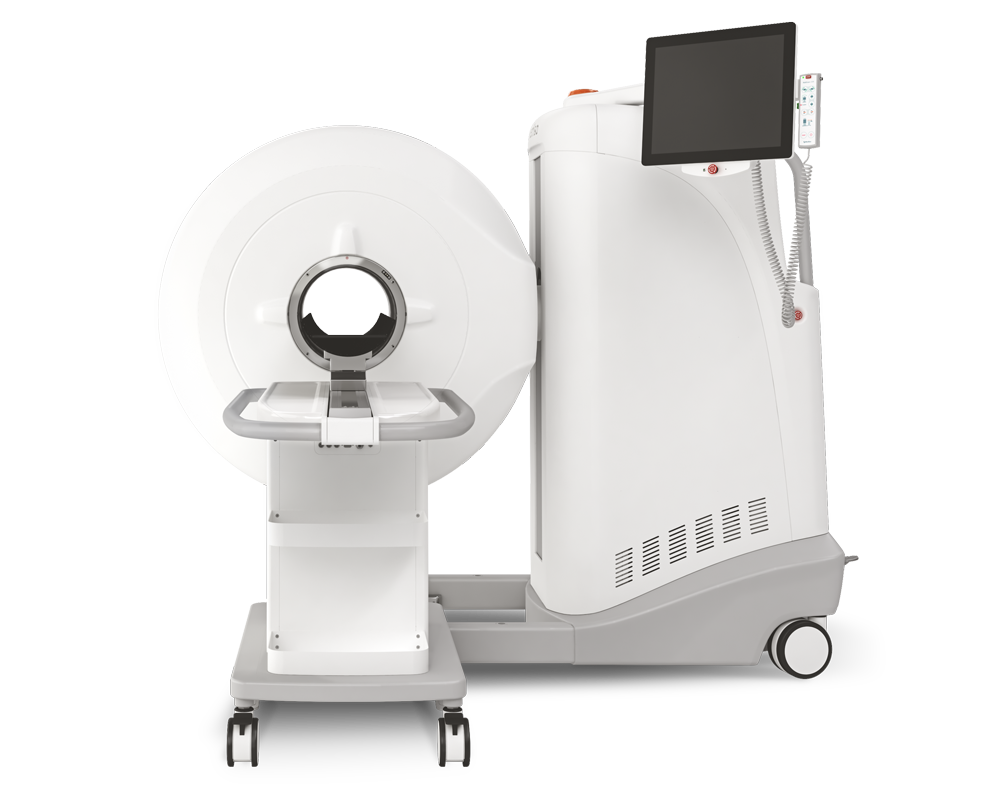
Results from MultiScan™ LFER PET/CT
- [18F]FDT allows direct monitoring of treatment that correlates with reduction in bacterial load
- [18F]FDT is also an Effective Mtb-Radiotracer in an ‘Old World’ Non-Human Primate TB Model
- [18F]FDT is non-toxic and [18F]FDT shows rapid clearance resulting in low background dose exposure
- [18F]FDT as a new, viable radiotracer for TB, suitable for Phase 1 trials

Figure 3.
Pulmonary [18F]FDT PET uptake into Tuberculous Lung Lesions is Reproducible within 90 min with Correlated Signal in Lesions with Higher Bacterial Loads.
A) [18F]FDT PET/CT scan of a naïve marmoset lung (administered 1 mCi) [18F]FDT and imaged 90 min post-injection (transverse view). B) [18F]FDT PET/CT scan of a representative, Mtb-infected marmoset lung showing lesions outlined in green and red (transverse view). The iliocostalis muscle was used for normalization of PET uptake (region outlined in blue). The target dose was 2.2 mCi/kg (~1 mCi). C) The [18F]FDT PET signal from Mtb-infected marmoset lungs is significantly higher than the signal from uninfected (control) marmoset lungs. The mean Standard uptake values (SUVbw) are represented as boxplots where the central bars represent the median. At least 4 lung regions of interest (ROI) were measured from 3 infected and 2 uninfected marmosets (p<0.0001). D) and E) Transverse images of [18F]FDT PET uptake scans for unblocked (D) and blocked (E) uptake in the same infected marmoset. [18F]FDT uptake was blocked by administering excess blocker, split into two administrations 1 h and 5 min prior to radiotracer administration. F) Reduction of [18F]FDT uptake in marmosets administered cold [19F]FDT blocker prior to being administered [18F]FDT (1.2 mCi) compared to when administered [18F]FDT alone. Animals (n = 22) were randomized as to the order of blocked scan; two days later the same animals were imaged with the treatments reversed. Uptake was measured in images collected 60 min and 90 min after probe administration. In both cases the animal receiving [19F]FDT blocking dose showed significantly lower accumulation of [18F]FDT into tubercular lesions. G) Comparison of [18F]FDT (1.0 mCi) lesion uptake (SUV/CMR) at 60, 90, and 120 mins post injection (lesions from two infected, antibiotic-treated marmosets) demonstrates that signal does not increase after 90 minutes. H) [18F]FDT uptake (SUV/CMR) is reproducible (r2 = 0.94), as illustrated by comparison of individual lesion SUV/CMR values from marmosets with imaging repeated two days apart (44 dpi and 46 dpi). I) [18F]FDT uptake into tubercular lesion tends to increase with higher mycobacterial loads (p = 0.001, ρ = 0.64). The total SUV of each lesion was compared with mycobacterial colony forming units measured (log CFU) in the lesions from two marmosets.
Wie können wir Ihnen behilflich sein?
Bitte kontaktieren Sie uns für technische Informationen und Unterstützung jeglicher Art in Zusammenhang mit unseren Entwicklungen und Produkten.
Kontaktformular